Phyre Home Retrieve Phyre Job Id
Emaill.a.kelley@imperial.ac.uk
DescriptionP38393
DateThu Jan 5 11:58:06 GMT 2012
Unique Job ID6c2165ff05641178

Summary 

Top model
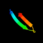
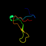
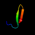
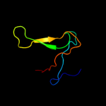
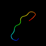
Image coloured by rainbow N → C terminus
Model (left) based on template c2d49A_
Top template information
PDB header:hydrolase
Chain: A: PDB Molecule:chitinase c;
PDBTitle: solution structure of the chitin-binding domain of2 streptomyces griseus chitinase c
Confidence and coverage
Confidence: 46.5% Coverage: 21%
15 residues ( 21% of your sequence) have been modelled with 46.5% confidence by the single highest scoring template.
You may wish to submit your sequence to Phyrealarm. This will automatically scan your sequence every week for new potential templates as they appear in the Phyre2 library.
Please note: You must be registered and logged in to use Phyrealarm.
3D viewing
Interactive 3D view in Jmol

Sequence analysis 

Secondary structure and disorder prediction 

   1........10.........20.........30.........40.........50.........60
Sequence  MIAHHFGTDEIPRQCVTPGDYVLHEGRTYIASANNIKKRKLYIRNLTTKTFITDRMIKVF
Secondary structure 


SS confidence 


Disorder  ???????


??
?


Disorder confidence 


   .........70...
Sequence  LGRDGLPVKAESW
Secondary structure 


SS confidence 


Disorder 


?
???????
Disorder confidence 


Confidence Key
High(9)                    Low (0)
?Disordered
Alpha helix
Beta strand

Domain analysis 

Hover over an aligned region to see model and summary info

Please note, only up to the top 20 hits are modelled to reduce computer load

RankAligned region

PDB 2d49 chain A

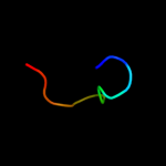
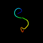
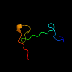
3D model

Region: 17 - 31
Aligned: 15
Modelled: 15
Confidence: 46.5%
Identity: 47%
PDB header:hydrolase
Chain: A: PDB Molecule:chitinase c;
PDBTitle: solution structure of the chitin-binding domain of2 streptomyces griseus chitinase c

Phyre2

PDB 1goi chain A domain 1

3D model

Region: 17 - 31
Aligned: 15
Modelled: 15
Confidence: 36.2%
Identity: 20%
Fold: WW domain-like
Superfamily: Carbohydrate binding domain
Family: Carbohydrate binding domain

Phyre2

PDB 1tie chain A

3D model

Region: 19 - 72
Aligned: 50
Modelled: 54
Confidence: 32.9%
Identity: 24%
Fold: beta-Trefoil
Superfamily: STI-like
Family: Kunitz (STI) inhibitors

Phyre2

PDB 2je6 chain I domain 2

3D model

Region: 12 - 59
Aligned: 46
Modelled: 48
Confidence: 30.1%
Identity: 22%
Fold: Barrel-sandwich hybrid
Superfamily: Ribosomal L27 protein-like
Family: ECR1 N-terminal domain-like

Phyre2

PDB 3bx1 chain C domain 1

3D model

Region: 19 - 69
Aligned: 51
Modelled: 51
Confidence: 21.0%
Identity: 20%
Fold: beta-Trefoil
Superfamily: STI-like
Family: Kunitz (STI) inhibitors

Phyre2

PDB 1r8o chain A

3D model

Region: 26 - 72
Aligned: 43
Modelled: 47
Confidence: 20.4%
Identity: 21%
PDB header:hydrolase inhibitor
Chain: A: PDB Molecule:kunitz trypsin inhibitor;
PDBTitle: crystal structure of an unusual kunitz-type trypsin inhibitor from2 copaifera langsdorffii seeds

Phyre2

PDB 2kca chain A

3D model

Region: 13 - 49
Aligned: 37
Modelled: 37
Confidence: 19.7%
Identity: 11%
PDB header:virus
Chain: A: PDB Molecule:bacteriophage spp1 complete nucleotide sequence;
PDBTitle: gp16

Phyre2

PDB 1aiw chain A

3D model

Region: 19 - 31
Aligned: 13
Modelled: 13
Confidence: 16.9%
Identity: 31%
Fold: WW domain-like
Superfamily: Carbohydrate binding domain
Family: Carbohydrate binding domain

Phyre2

PDB 1ed7 chain A

3D model

Region: 14 - 31
Aligned: 18
Modelled: 18
Confidence: 16.8%
Identity: 33%
Fold: WW domain-like
Superfamily: Carbohydrate binding domain
Family: Carbohydrate binding domain

Phyre2

PDB 1eyl chain A

3D model

Region: 19 - 73
Aligned: 55
Modelled: 55
Confidence: 15.3%
Identity: 13%
Fold: beta-Trefoil
Superfamily: STI-like
Family: Kunitz (STI) inhibitors

Phyre2

PDB 2ba0 chain A domain 2

3D model

Region: 13 - 59
Aligned: 45
Modelled: 47
Confidence: 15.1%
Identity: 18%
Fold: Barrel-sandwich hybrid
Superfamily: Ribosomal L27 protein-like
Family: ECR1 N-terminal domain-like

Phyre2

PDB 2eif chain A

3D model

Region: 7 - 31
Aligned: 25
Modelled: 25
Confidence: 14.1%
Identity: 24%
PDB header:gene regulation
Chain: A: PDB Molecule:protein (eukaryotic translation initiation factor 5a);
PDBTitle: eukaryotic translation initiation factor 5a from methanococcus2 jannaschii

Phyre2

PDB 3iir chain A

3D model

Region: 19 - 73
Aligned: 55
Modelled: 55
Confidence: 13.9%
Identity: 13%
PDB header:hydrolase inhibitor
Chain: A: PDB Molecule:trypsin inhibitor;
PDBTitle: crystal structure of miraculin like protein from seeds of murraya2 koenigii

Phyre2

PDB 2qn4 chain B

3D model

Region: 19 - 69
Aligned: 46
Modelled: 51
Confidence: 11.5%
Identity: 24%
PDB header:hydrolase inhibitor
Chain: B: PDB Molecule:alpha-amylase/subtilisin inhibitor;
PDBTitle: structure and function study of rice bifunctional alpha-2 amylase/subtilisin inhibitor from oryza sativa

Phyre2

PDB 1j83 chain A

3D model

Region: 16 - 31
Aligned: 16
Modelled: 16
Confidence: 11.5%
Identity: 31%
Fold: Galactose-binding domain-like
Superfamily: Galactose-binding domain-like
Family: Family 17 carbohydrate binding module, CBM17

Phyre2

PDB 3flo chain G

3D model

Region: 4 - 52
Aligned: 49
Modelled: 49
Confidence: 10.9%
Identity: 16%
PDB header:transferase
Chain: G: PDB Molecule:dna polymerase alpha subunit b;
PDBTitle: crystal structure of the carboxyl-terminal domain of yeast2 dna polymerase alpha in complex with its b subunit

Phyre2

PDB 1tdh chain A domain 3

3D model

Region: 17 - 28
Aligned: 12
Modelled: 12
Confidence: 10.5%
Identity: 42%
Fold: Glucocorticoid receptor-like (DNA-binding domain)
Superfamily: Glucocorticoid receptor-like (DNA-binding domain)
Family: C-terminal, Zn-finger domain of MutM-like DNA repair proteins

Phyre2

PDB 2juf chain A

3D model

Region: 12 - 23
Aligned: 12
Modelled: 12
Confidence: 10.4%
Identity: 33%
PDB header:gene regulation
Chain: A: PDB Molecule:p53-associated parkin-like cytoplasmic protein;
PDBTitle: nmr solution structure of parc cph domain. nesg target2 hr3443b/sgc-toronto

Phyre2

PDB 2jng chain A domain 1

3D model

Region: 12 - 23
Aligned: 12
Modelled: 12
Confidence: 9.9%
Identity: 33%
Fold: SH3-like barrel
Superfamily: Tudor/PWWP/MBT
Family: CPH domain

Phyre2

PDB 1lkt chain A

3D model

Region: 27 - 63
Aligned: 33
Modelled: 37
Confidence: 9.3%
Identity: 33%
Fold: Head-binding domain of phage P22 tailspike protein
Superfamily: Head-binding domain of phage P22 tailspike protein
Family: Head-binding domain of phage P22 tailspike protein

Phyre2
1

c2d49A_
2

d1goia1
3

d1tiea_
4

d2je6i2
5

d3bx1c1
6

c1r8oA_
7

c2kcaA_
8

d1aiwa_
9

d1ed7a_
10

d1eyla_
11

d2ba0a2
12

c2eifA_
13

c3iirA_
14

c2qn4B_
15

d1j83a_
16

c3floG_
17

d1tdha3
18

c2jufA_
19

d2jnga1
20

d1lkta_
21


22


23


24


25


26


27


28


29


Detailed template information 

#
Template Alignment Coverage3D Model Confidence
% i.d. Template Information
1c2d49A_


46.5 47 PDB header:hydrolase
Chain: A: PDB Molecule:chitinase c;
PDBTitle: solution structure of the chitin-binding domain of2 streptomyces griseus chitinase c
2d1goia1


36.2 20 Fold:WW domain-like
Superfamily:Carbohydrate binding domain
Family:Carbohydrate binding domain
3d1tiea_


32.9 24 Fold:beta-Trefoil
Superfamily:STI-like
Family:Kunitz (STI) inhibitors
4d2je6i2


30.1 22 Fold:Barrel-sandwich hybrid
Superfamily:Ribosomal L27 protein-like
Family:ECR1 N-terminal domain-like
5d3bx1c1


21.0 20 Fold:beta-Trefoil
Superfamily:STI-like
Family:Kunitz (STI) inhibitors
6c1r8oA_


20.4 21 PDB header:hydrolase inhibitor
Chain: A: PDB Molecule:kunitz trypsin inhibitor;
PDBTitle: crystal structure of an unusual kunitz-type trypsin inhibitor from2 copaifera langsdorffii seeds
7c2kcaA_


19.7 11 PDB header:virus
Chain: A: PDB Molecule:bacteriophage spp1 complete nucleotide sequence;
PDBTitle: gp16
8d1aiwa_


16.9 31 Fold:WW domain-like
Superfamily:Carbohydrate binding domain
Family:Carbohydrate binding domain
9d1ed7a_


16.8 33 Fold:WW domain-like
Superfamily:Carbohydrate binding domain
Family:Carbohydrate binding domain
10d1eyla_


15.3 13 Fold:beta-Trefoil
Superfamily:STI-like
Family:Kunitz (STI) inhibitors
11d2ba0a2


15.1 18 Fold:Barrel-sandwich hybrid
Superfamily:Ribosomal L27 protein-like
Family:ECR1 N-terminal domain-like
12c2eifA_


14.1 24 PDB header:gene regulation
Chain: A: PDB Molecule:protein (eukaryotic translation initiation factor 5a);
PDBTitle: eukaryotic translation initiation factor 5a from methanococcus2 jannaschii
13c3iirA_


13.9 13 PDB header:hydrolase inhibitor
Chain: A: PDB Molecule:trypsin inhibitor;
PDBTitle: crystal structure of miraculin like protein from seeds of murraya2 koenigii
14c2qn4B_


11.5 24 PDB header:hydrolase inhibitor
Chain: B: PDB Molecule:alpha-amylase/subtilisin inhibitor;
PDBTitle: structure and function study of rice bifunctional alpha-2 amylase/subtilisin inhibitor from oryza sativa
15d1j83a_


11.5 31 Fold:Galactose-binding domain-like
Superfamily:Galactose-binding domain-like
Family:Family 17 carbohydrate binding module, CBM17
16c3floG_


10.9 16 PDB header:transferase
Chain: G: PDB Molecule:dna polymerase alpha subunit b;
PDBTitle: crystal structure of the carboxyl-terminal domain of yeast2 dna polymerase alpha in complex with its b subunit
17d1tdha3


10.5 42 Fold:Glucocorticoid receptor-like (DNA-binding domain)
Superfamily:Glucocorticoid receptor-like (DNA-binding domain)
Family:C-terminal, Zn-finger domain of MutM-like DNA repair proteins
18c2jufA_


10.4 33 PDB header:gene regulation
Chain: A: PDB Molecule:p53-associated parkin-like cytoplasmic protein;
PDBTitle: nmr solution structure of parc cph domain. nesg target2 hr3443b/sgc-toronto
19d2jnga1


9.9 33 Fold:SH3-like barrel
Superfamily:Tudor/PWWP/MBT
Family:CPH domain
20d1lkta_


9.3 33 Fold:Head-binding domain of phage P22 tailspike protein
Superfamily:Head-binding domain of phage P22 tailspike protein
Family:Head-binding domain of phage P22 tailspike protein
21c1fmeA_


not modelled 7.9 33 PDB header:de novo protein
Chain: A: PDB Molecule:fsd-ey peptide;
PDBTitle: solution structure of fsd-ey, a novel peptide assuming a2 beta-beta-alpha fold
22c3s9xA_


not modelled 7.3 33 PDB header:structural genomics, unknown function
Chain: A: PDB Molecule:asch domain;
PDBTitle: high resolution crystal structure of asch domain from lactobacillus2 crispatus jv v101
23d1t62a_


not modelled 7.2 17 Fold:PUA domain-like
Superfamily:PUA domain-like
Family:Hypothetical protein EF3133
24c1bkbA_


not modelled 7.1 20 PDB header:translation
Chain: A: PDB Molecule:translation initiation factor 5a;
PDBTitle: initiation factor 5a from archebacterium pyrobaculum2 aerophilum
25c3hksB_


not modelled 7.1 13 PDB header:translation, rna binding protein
Chain: B: PDB Molecule:eukaryotic translation initiation factor 5a-2;
PDBTitle: crystal structure of eukaryotic translation initiation2 factor eif-5a2 from arabidopsis thaliana
26c2l23A_


not modelled 7.0 24 PDB header:transcription
Chain: A: PDB Molecule:mediator of rna polymerase ii transcription subunit 25;
PDBTitle: nmr structure of the acid (activator interacting domain) of the human2 mediator med25 protein
27d2nn6h2


not modelled 6.9 16 Fold:Barrel-sandwich hybrid
Superfamily:Ribosomal L27 protein-like
Family:ECR1 N-terminal domain-like
28d1a1xa_


not modelled 6.6 20 Fold:Oncogene products
Superfamily:Oncogene products
Family:Oncogene products
29d1wbaa_


not modelled 5.3 9 Fold:beta-Trefoil
Superfamily:STI-like
Family:Kunitz (STI) inhibitors

Binding site prediction 

Due to computational demand, binding site predictions are not run for batch jobs

If you want to predict binding sites, please manually submit your model of choice to 3DLigandSite


Phyre is for academic use only

Please cite: Protein structure prediction on the web: a case study using the Phyre server
Kelley LA and Sternberg MJE. Nature Protocols 4, 363 - 371 (2009) [pdf] [Import into BibTeX]
 
If you use the binding site predictions from 3DLigandSite, please also cite:
3DLigandSite: predicting ligand-binding sites using similar structures.
Wass MN, Kelley LA and Sternberg MJ Nucleic Acids Research 38, W469-73 (2010) [PubMed]
 
© Structural Bioinformatics Group
Imperial College London
Lawrence Kelley, Benjamin Jefferys
Disclaimer
Terms and Conditions
Component software
Template detection: HHpred 1.51
Secondary structure prediction: Psi-pred 2.5
Disorder prediction: Disopred 2.4
Transmembrane prediction: Memsat_SVM
Multi-template modelling and ab initio: Poing 1.0