Phyre Home Retrieve Phyre Job Id
Emaill.a.kelley@imperial.ac.uk
DescriptionP0AC16
DateThu Jan 5 11:16:54 GMT 2012
Unique Job ID6bf811766217f25c

Summary 

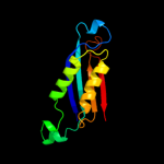
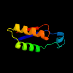
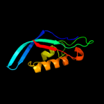
Top model
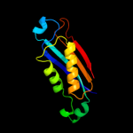
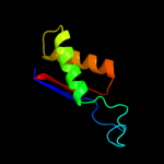
Image coloured by rainbow N → C terminus
Model (left) based on template c2o90A_
Top template information
PDB header:lyase
Chain: A: PDB Molecule:dihydroneopterin aldolase;
PDBTitle: atomic resolution crystal structure of e.coli2 dihydroneopterin aldolase in complex with neopterin
Confidence and coverage
Confidence:100.0% Coverage: 94%
115 residues ( 94% of your sequence) have been modelled with 100.0% confidence by the single highest scoring template.
3D viewing
Interactive 3D view in Jmol

Sequence analysis 

Secondary structure and disorder prediction 

   1........10.........20.........30.........40.........50.........60
Sequence  MDIVFIEQLSVITTIGVYDWEQTIEQKLVFDIEMAWDNRKAAKSDDVADCLSYADIAETV
Secondary structure 


SS confidence 


Disorder  ??


Disorder confidence 


   .........70.........80.........90.........100.........110.........120
Sequence  VSHVEGARFALVERVAEEVAELLLARFNSPWVRIKLSKPGAVARAANVGVIIERGNNLKE
Secondary structure 


SS confidence 


Disorder 


??????
Disorder confidence 


   ..
Sequence  NN
Secondary structure 

SS confidence 

Disorder  ??
Disorder confidence 


Confidence Key
High(9)                    Low (0)
?Disordered
Alpha helix
Beta strand

Domain analysis 

Hover over an aligned region to see model and summary info

Please note, only up to the top 20 hits are modelled to reduce computer load

RankAligned region

PDB 2o90 chain A

3D model

Region: 1 - 115
Aligned: 115
Modelled: 115
Confidence: 100.0%
Identity: 100%
PDB header:lyase
Chain: A: PDB Molecule:dihydroneopterin aldolase;
PDBTitle: atomic resolution crystal structure of e.coli2 dihydroneopterin aldolase in complex with neopterin

Phyre2

PDB 1sql chain N

3D model

Region: 2 - 117
Aligned: 116
Modelled: 116
Confidence: 100.0%
Identity: 34%
PDB header:lyase
Chain: N: PDB Molecule:dihydroneopterin aldolase;
PDBTitle: crystal structure of 7,8-dihydroneopterin aldolase in2 complex with guanine

Phyre2

PDB 1b9l chain A

3D model

Region: 1 - 116
Aligned: 116
Modelled: 116
Confidence: 100.0%
Identity: 24%
Fold: T-fold
Superfamily: Tetrahydrobiopterin biosynthesis enzymes-like
Family: DHN aldolase/epimerase

Phyre2

PDB 1sql chain A

3D model

Region: 2 - 116
Aligned: 115
Modelled: 115
Confidence: 100.0%
Identity: 34%
Fold: T-fold
Superfamily: Tetrahydrobiopterin biosynthesis enzymes-like
Family: DHN aldolase/epimerase

Phyre2

PDB 1nbu chain A

3D model

Region: 2 - 116
Aligned: 115
Modelled: 115
Confidence: 100.0%
Identity: 31%
Fold: T-fold
Superfamily: Tetrahydrobiopterin biosynthesis enzymes-like
Family: DHN aldolase/epimerase

Phyre2

PDB 1dhn chain A

3D model

Region: 1 - 117
Aligned: 117
Modelled: 117
Confidence: 100.0%
Identity: 26%
Fold: T-fold
Superfamily: Tetrahydrobiopterin biosynthesis enzymes-like
Family: DHN aldolase/epimerase

Phyre2

PDB 2cg8 chain B

3D model

Region: 1 - 118
Aligned: 114
Modelled: 114
Confidence: 100.0%
Identity: 23%
PDB header:lyase/transferase
Chain: B: PDB Molecule:dihydroneopterin aldolase 6-hydroxymethyl-7,8-
PDBTitle: the bifunctional dihydroneopterin aldolase 6-hydroxymethyl-2 7,8-dihydropterin synthase from streptococcus pneumoniae

Phyre2

PDB 2iba chain A domain 1

3D model

Region: 28 - 97
Aligned: 70
Modelled: 70
Confidence: 94.9%
Identity: 7%
Fold: T-fold
Superfamily: Tetrahydrobiopterin biosynthesis enzymes-like
Family: Urate oxidase (uricase)

Phyre2

PDB 1j2g chain A domain 1

3D model

Region: 28 - 97
Aligned: 70
Modelled: 70
Confidence: 93.8%
Identity: 11%
Fold: T-fold
Superfamily: Tetrahydrobiopterin biosynthesis enzymes-like
Family: Urate oxidase (uricase)

Phyre2

PDB 2yzc chain A domain 1

3D model

Region: 28 - 97
Aligned: 69
Modelled: 70
Confidence: 93.8%
Identity: 17%
Fold: T-fold
Superfamily: Tetrahydrobiopterin biosynthesis enzymes-like
Family: Urate oxidase (uricase)

Phyre2

PDB 1r56 chain H

3D model

Region: 28 - 97
Aligned: 70
Modelled: 70
Confidence: 92.7%
Identity: 7%
PDB header:oxidoreductase
Chain: H: PDB Molecule:uricase;
PDBTitle: uncomplexed urate oxidase from aspergillus flavus

Phyre2

PDB 2yzb chain A

3D model

Region: 28 - 97
Aligned: 69
Modelled: 70
Confidence: 92.0%
Identity: 17%
PDB header:oxidoreductase
Chain: A: PDB Molecule:uricase;
PDBTitle: crystal structure of uricase from arthrobacter globiformis2 in complex with uric acid (substrate)

Phyre2

PDB 1j2g chain C

3D model

Region: 28 - 97
Aligned: 70
Modelled: 70
Confidence: 90.1%
Identity: 11%
PDB header:oxidoreductase
Chain: C: PDB Molecule:uricase;
PDBTitle: crystal structure of urate oxidase from bacillus sp. tb-90 co-2 crystallized with 8-azaxanthine

Phyre2

PDB 2a0s chain A domain 1

3D model

Region: 1 - 97
Aligned: 91
Modelled: 97
Confidence: 76.3%
Identity: 15%
Fold: T-fold
Superfamily: Tetrahydrobiopterin biosynthesis enzymes-like
Family: 6-pyruvoyl tetrahydropterin synthase

Phyre2

PDB 2r5r chain A

3D model

Region: 1 - 97
Aligned: 84
Modelled: 95
Confidence: 69.9%
Identity: 17%
PDB header:structural genomics, unknown function
Chain: A: PDB Molecule:upf0343 protein ne1163;
PDBTitle: the crystal structure of duf198 from nitrosomonas europaea2 atcc 19718

Phyre2

PDB 1y13 chain A

3D model

Region: 3 - 97
Aligned: 89
Modelled: 95
Confidence: 55.6%
Identity: 18%
Fold: T-fold
Superfamily: Tetrahydrobiopterin biosynthesis enzymes-like
Family: 6-pyruvoyl tetrahydropterin synthase

Phyre2

PDB 3d2o chain B

3D model

Region: 1 - 97
Aligned: 83
Modelled: 93
Confidence: 53.5%
Identity: 24%
PDB header:hydrolase, biosynthetic protein
Chain: B: PDB Molecule:upf0343 protein ngo0387;
PDBTitle: crystal structure of manganese-metallated gtp cyclohydrolase2 type ib

Phyre2

PDB 2dj6 chain B

3D model

Region: 1 - 97
Aligned: 90
Modelled: 97
Confidence: 32.7%
Identity: 12%
PDB header:lyase
Chain: B: PDB Molecule:hypothetical protein ph0634;
PDBTitle: crystal structure of 6-pyruvoyl tetrahydrobiopterin synthase from2 pyrococcus horikoshii ot3

Phyre2

PDB 1j2g chain A domain 2

3D model

Region: 29 - 96
Aligned: 68
Modelled: 68
Confidence: 25.6%
Identity: 12%
Fold: T-fold
Superfamily: Tetrahydrobiopterin biosynthesis enzymes-like
Family: Urate oxidase (uricase)

Phyre2

PDB 1a6q chain A domain 2

3D model

Region: 44 - 103
Aligned: 58
Modelled: 60
Confidence: 21.7%
Identity: 19%
Fold: PP2C-like
Superfamily: PP2C-like
Family: PP2C-like

Phyre2
1

c2o90A_
2

c1sqlN_
3

d1b9la_
4

d1sqla_
5

d1nbua_
6

d1dhna_
7

c2cg8B_
8

d2ibaa1
9

d1j2ga1
10

d2yzca1
11

c1r56H_
12

c2yzbA_
13

c1j2gC_
14

d2a0sa1
15

c2r5rA_
16

d1y13a_
17

c3d2oB_
18

c2dj6B_
19

d1j2ga2
20

d1a6qa2
21


22


23


24


25


26


27


28


29


30


31


32


Detailed template information 

#
Template Alignment Coverage3D Model Confidence
% i.d. Template Information
1c2o90A_


100.0 100 PDB header:lyase
Chain: A: PDB Molecule:dihydroneopterin aldolase;
PDBTitle: atomic resolution crystal structure of e.coli2 dihydroneopterin aldolase in complex with neopterin
2c1sqlN_


100.0 34 PDB header:lyase
Chain: N: PDB Molecule:dihydroneopterin aldolase;
PDBTitle: crystal structure of 7,8-dihydroneopterin aldolase in2 complex with guanine
3d1b9la_


100.0 24 Fold:T-fold
Superfamily:Tetrahydrobiopterin biosynthesis enzymes-like
Family:DHN aldolase/epimerase
4d1sqla_


100.0 34 Fold:T-fold
Superfamily:Tetrahydrobiopterin biosynthesis enzymes-like
Family:DHN aldolase/epimerase
5d1nbua_


100.0 31 Fold:T-fold
Superfamily:Tetrahydrobiopterin biosynthesis enzymes-like
Family:DHN aldolase/epimerase
6d1dhna_


100.0 26 Fold:T-fold
Superfamily:Tetrahydrobiopterin biosynthesis enzymes-like
Family:DHN aldolase/epimerase
7c2cg8B_


100.0 23 PDB header:lyase/transferase
Chain: B: PDB Molecule:dihydroneopterin aldolase 6-hydroxymethyl-7,8-
PDBTitle: the bifunctional dihydroneopterin aldolase 6-hydroxymethyl-2 7,8-dihydropterin synthase from streptococcus pneumoniae
8d2ibaa1


94.9 7 Fold:T-fold
Superfamily:Tetrahydrobiopterin biosynthesis enzymes-like
Family:Urate oxidase (uricase)
9d1j2ga1


93.8 11 Fold:T-fold
Superfamily:Tetrahydrobiopterin biosynthesis enzymes-like
Family:Urate oxidase (uricase)
10d2yzca1


93.8 17 Fold:T-fold
Superfamily:Tetrahydrobiopterin biosynthesis enzymes-like
Family:Urate oxidase (uricase)
11c1r56H_


92.7 7 PDB header:oxidoreductase
Chain: H: PDB Molecule:uricase;
PDBTitle: uncomplexed urate oxidase from aspergillus flavus
12c2yzbA_


92.0 17 PDB header:oxidoreductase
Chain: A: PDB Molecule:uricase;
PDBTitle: crystal structure of uricase from arthrobacter globiformis2 in complex with uric acid (substrate)
13c1j2gC_


90.1 11 PDB header:oxidoreductase
Chain: C: PDB Molecule:uricase;
PDBTitle: crystal structure of urate oxidase from bacillus sp. tb-90 co-2 crystallized with 8-azaxanthine
14d2a0sa1


76.3 15 Fold:T-fold
Superfamily:Tetrahydrobiopterin biosynthesis enzymes-like
Family:6-pyruvoyl tetrahydropterin synthase
15c2r5rA_


69.9 17 PDB header:structural genomics, unknown function
Chain: A: PDB Molecule:upf0343 protein ne1163;
PDBTitle: the crystal structure of duf198 from nitrosomonas europaea2 atcc 19718
16d1y13a_


55.6 18 Fold:T-fold
Superfamily:Tetrahydrobiopterin biosynthesis enzymes-like
Family:6-pyruvoyl tetrahydropterin synthase
17c3d2oB_


53.5 24 PDB header:hydrolase, biosynthetic protein
Chain: B: PDB Molecule:upf0343 protein ngo0387;
PDBTitle: crystal structure of manganese-metallated gtp cyclohydrolase2 type ib
18c2dj6B_


32.7 12 PDB header:lyase
Chain: B: PDB Molecule:hypothetical protein ph0634;
PDBTitle: crystal structure of 6-pyruvoyl tetrahydrobiopterin synthase from2 pyrococcus horikoshii ot3
19d1j2ga2


25.6 12 Fold:T-fold
Superfamily:Tetrahydrobiopterin biosynthesis enzymes-like
Family:Urate oxidase (uricase)
20d1a6qa2


21.7 19 Fold:PP2C-like
Superfamily:PP2C-like
Family:PP2C-like
21c3gdzA_


not modelled 15.6 11 PDB header:ligase
Chain: A: PDB Molecule:arginyl-trna synthetase;
PDBTitle: crystal structure of arginyl-trna synthetase from klebsiella2 pneumoniae subsp. pneumoniae
22c2i44A_


not modelled 13.8 16 PDB header:hydrolase
Chain: A: PDB Molecule:serine-threonine phosphatase 2c;
PDBTitle: crystal structure of serine-threonine phosphatase 2c from2 toxoplasma gondii
23d1ghha_


not modelled 10.6 21 Fold:DNA damage-inducible protein DinI
Superfamily:DNA damage-inducible protein DinI
Family:DNA damage-inducible protein DinI
24d1a04a2


not modelled 9.8 16 Fold:Flavodoxin-like
Superfamily:CheY-like
Family:CheY-related
25d2yzca2


not modelled 9.2 9 Fold:T-fold
Superfamily:Tetrahydrobiopterin biosynthesis enzymes-like
Family:Urate oxidase (uricase)
26d1wura1


not modelled 8.3 20 Fold:T-fold
Superfamily:Tetrahydrobiopterin biosynthesis enzymes-like
Family:GTP cyclohydrolase I
27c1wm9D_


not modelled 8.3 20 PDB header:hydrolase
Chain: D: PDB Molecule:gtp cyclohydrolase i;
PDBTitle: structure of gtp cyclohydrolase i from thermus thermophilus hb8
28d1wpla_


not modelled 7.6 13 Fold:T-fold
Superfamily:Tetrahydrobiopterin biosynthesis enzymes-like
Family:GTP cyclohydrolase I
29c1is7F_


not modelled 7.6 13 PDB header:hydrolase/protein binding
Chain: F: PDB Molecule:gtp cyclohydrolase i;
PDBTitle: crystal structure of rat gtpchi/gfrp stimulatory complex
30d2ibaa2


not modelled 7.5 25 Fold:T-fold
Superfamily:Tetrahydrobiopterin biosynthesis enzymes-like
Family:Urate oxidase (uricase)
31d1a8ra_


not modelled 6.9 15 Fold:T-fold
Superfamily:Tetrahydrobiopterin biosynthesis enzymes-like
Family:GTP cyclohydrolase I
32c2zw2B_


not modelled 6.7 12 PDB header:ligase
Chain: B: PDB Molecule:putative uncharacterized protein sts178;
PDBTitle: crystal structure of formylglycinamide ribonucleotide amidotransferase2 iii from sulfolobus tokodaii (stpurs)

Binding site prediction 

Due to computational demand, binding site predictions are not run for batch jobs

If you want to predict binding sites, please manually submit your model of choice to 3DLigandSite


Phyre is for academic use only

Please cite: Protein structure prediction on the web: a case study using the Phyre server
Kelley LA and Sternberg MJE. Nature Protocols 4, 363 - 371 (2009) [pdf] [Import into BibTeX]
 
If you use the binding site predictions from 3DLigandSite, please also cite:
3DLigandSite: predicting ligand-binding sites using similar structures.
Wass MN, Kelley LA and Sternberg MJ Nucleic Acids Research 38, W469-73 (2010) [PubMed]
 
© Structural Bioinformatics Group
Imperial College London
Lawrence Kelley, Benjamin Jefferys
Disclaimer
Terms and Conditions
Component software
Template detection: HHpred 1.51
Secondary structure prediction: Psi-pred 2.5
Disorder prediction: Disopred 2.4
Transmembrane prediction: Memsat_SVM
Multi-template modelling and ab initio: Poing 1.0