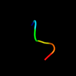
| 1 |
|
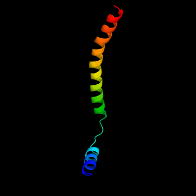
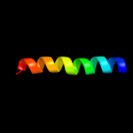
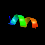
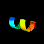
PDB 1rh5 chain B
Region: 1 - 55
Aligned: 55
Modelled: 55
Confidence: 12.1%
Identity: 15%
Fold: Single transmembrane helix
Superfamily: Preprotein translocase SecE subunit
Family: Preprotein translocase SecE subunit
Phyre2
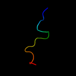
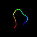
| 2 |
|
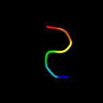
PDB 2dyo chain B
Region: 70 - 79
Aligned: 10
Modelled: 10
Confidence: 11.2%
Identity: 40%
PDB header:protein turnover/protein turnover
Chain: B: PDB Molecule:autophagy protein 16;
PDBTitle: the crystal structure of saccharomyces cerevisiae atg5-2 atg16(1-57) complex
Phyre2
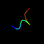
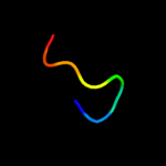
| 3 |
|
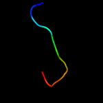
PDB 2l2t chain A
Region: 51 - 74
Aligned: 24
Modelled: 24
Confidence: 10.7%
Identity: 21%
PDB header:membrane protein
Chain: A: PDB Molecule:receptor tyrosine-protein kinase erbb-4;
PDBTitle: solution nmr structure of the erbb4 dimeric membrane domain
Phyre2
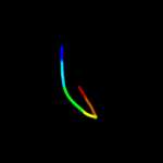
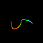
| 4 |
|
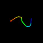
PDB 2f22 chain A domain 1
Region: 2 - 20
Aligned: 19
Modelled: 19
Confidence: 9.6%
Identity: 37%
Fold: DinB/YfiT-like putative metalloenzymes
Superfamily: DinB/YfiT-like putative metalloenzymes
Family: DinB-like
Phyre2
| 5 |
|
PDB 1bzg chain A
Region: 75 - 81
Aligned: 7
Modelled: 7
Confidence: 8.9%
Identity: 57%
PDB header:hormone
Chain: A: PDB Molecule:parathyroid hormone-related protein;
PDBTitle: the solution structure of human parathyroid hormone-related2 protein (1-34) in near-physiological solution, nmr, 303 structures
Phyre2
| 6 |
|
PDB 1zcz chain A
Region: 112 - 117
Aligned: 6
Modelled: 6
Confidence: 8.8%
Identity: 50%
PDB header:transferase/hydrolase
Chain: A: PDB Molecule:bifunctional purine biosynthesis protein purh;
PDBTitle: crystal structure of phosphoribosylaminoimidazolecarboxamide2 formyltransferase / imp cyclohydrolase (tm1249) from thermotoga3 maritima at 1.88 a resolution
Phyre2
| 7 |
|
PDB 3skd chain A
Region: 75 - 80
Aligned: 6
Modelled: 6
Confidence: 8.6%
Identity: 50%
PDB header:hydrolase
Chain: A: PDB Molecule:putative uncharacterized protein tthb187;
PDBTitle: crystal structure of the thermus thermophilus cas3 hd domain in the2 presence of ni2+
Phyre2
| 8 |
|
PDB 2lkj chain A
Region: 114 - 121
Aligned: 8
Modelled: 8
Confidence: 7.6%
Identity: 50%
PDB header:cell adhesion
Chain: A: PDB Molecule:integrin alpha-m;
PDBTitle: structures and interaction analyses of the integrin alpha-m beta-22 cytoplasmic tails
Phyre2
| 9 |
|
PDB 1lir chain A
Region: 73 - 80
Aligned: 8
Modelled: 8
Confidence: 6.8%
Identity: 63%
Fold: Knottins (small inhibitors, toxins, lectins)
Superfamily: Scorpion toxin-like
Family: Short-chain scorpion toxins
Phyre2
| 10 |
|
PDB 2ogi chain A
Region: 75 - 80
Aligned: 6
Modelled: 6
Confidence: 6.7%
Identity: 50%
PDB header:hydrolase
Chain: A: PDB Molecule:hypothetical protein sag1661;
PDBTitle: crystal structure of a putative metal dependent phosphohydrolase2 (sag1661) from streptococcus agalactiae serogroup v at 1.85 a3 resolution
Phyre2
| 11 |
|
PDB 3zrk chain Y
Region: 67 - 76
Aligned: 10
Modelled: 10
Confidence: 6.4%
Identity: 40%
PDB header:transferase/peptide
Chain: Y: PDB Molecule:proto-oncogene frat1;
PDBTitle: identification of 2-(4-pyridyl)thienopyridinones as gsk-3beta2 inhibitors
Phyre2
| 12 |
|
PDB 2o08 chain B
Region: 75 - 80
Aligned: 6
Modelled: 6
Confidence: 6.3%
Identity: 33%
PDB header:hydrolase
Chain: B: PDB Molecule:bh1327 protein;
PDBTitle: crystal structure of a putative hd superfamily hydrolase (bh1327) from2 bacillus halodurans at 1.90 a resolution
Phyre2
| 13 |
|
PDB 2rf9 chain D
Region: 108 - 119
Aligned: 12
Modelled: 12
Confidence: 6.0%
Identity: 50%
PDB header:transferase
Chain: D: PDB Molecule:erbb receptor feedback inhibitor 1;
PDBTitle: crystal structure of the complex between the egfr kinase2 domain and a mig6 peptide
Phyre2
| 14 |
|
PDB 3ccg chain A
Region: 75 - 80
Aligned: 6
Modelled: 6
Confidence: 5.8%
Identity: 33%
PDB header:hydrolase
Chain: A: PDB Molecule:hd superfamily hydrolase;
PDBTitle: crystal structure of predicted hd superfamily hydrolase involved in2 nad metabolism (np_347894.1) from clostridium acetobutylicum at 1.503 a resolution
Phyre2
| 15 |
|
PDB 1gng chain X
Region: 67 - 76
Aligned: 10
Modelled: 10
Confidence: 5.7%
Identity: 40%
PDB header:transferase
Chain: X: PDB Molecule:frattide;
PDBTitle: glycogen synthase kinase-3 beta (gsk3) complex with frattide2 peptide
Phyre2
| 16 |
|
PDB 1thz chain A
Region: 112 - 121
Aligned: 10
Modelled: 10
Confidence: 5.6%
Identity: 40%
PDB header:transferase, hydrolase
Chain: A: PDB Molecule:bifunctional purine biosynthesis protein purh;
PDBTitle: crystal structure of avian aicar transformylase in complex2 with a novel inhibitor identified by virtual ligand3 screening
Phyre2