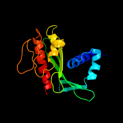
Phyre Home Retrieve Phyre Job Id
Emaill.a.kelley@imperial.ac.uk
DescriptionP77285
DateThu Jan 5 12:27:13 GMT 2012
Unique Job ID6a5e84c5a45feaee

Summary 

Top model
Image coloured by rainbow N → C terminus
Model (left) based on template c3lwxA_
Top template information
PDB header:oxidoreductase
Chain: A: PDB Molecule:nadh:ubiquinone oxidoreductase, na translocating,
PDBTitle: crystal structure of na(+)-translocating nadh-quinone2 reductase subunit c (yp_001302508.1) from parabacteroides3 distasonis atcc 8503 at 1.10 a resolution
Confidence and coverage
Confidence:100.0% Coverage: 79%
162 residues ( 79% of your sequence) have been modelled with 100.0% confidence by the single highest scoring template.
3D viewing
Interactive 3D view in Jmol

Sequence analysis 

Secondary structure and disorder prediction 

   1........10.........20.........30.........40.........50.........60
Sequence  MLKTIRKHGITLALFAAGSTGLTAAINQMTKTTIAEQASLQQKALFDQVLPAERYNNALA
Secondary structure 


SS confidence 


Disorder  ??


???


Disorder confidence 


   .........70.........80.........90.........100.........110.........120
Sequence  QSCYLVTAPELGKGEHRVYIAKQDDKPVAAVLEATAPDGYSGAIQLLVGADFNGTVLGTR
Secondary structure 


SS confidence 


Disorder 
?????????????


?


Disorder confidence 


   .........130.........140.........150.........160.........170.........180
Sequence  VTEHHETPGLGDKIELRLSDWITHFAGKKISGADDAHWAVKKDGGDFDQFTGATITPRAV
Secondary structure 


SS confidence 


Disorder 


???
????????
??????


Disorder confidence 


   .........190.........200......
Sequence  VNAVKRAGLYAQTLPAQLSQLPACGE
Secondary structure 


SS confidence 


Disorder 


???????
Disorder confidence 


Confidence Key
High(9)                    Low (0)
?Disordered
Alpha helix
Beta strand

Domain analysis 

Hover over an aligned region to see model and summary info

Please note, only up to the top 20 hits are modelled to reduce computer load

RankAligned region

PDB 3lwx chain A

3D model

Region: 28 - 193
Aligned: 162
Modelled: 166
Confidence: 100.0%
Identity: 19%
PDB header:oxidoreductase
Chain: A: PDB Molecule:nadh:ubiquinone oxidoreductase, na translocating,
PDBTitle: crystal structure of na(+)-translocating nadh-quinone2 reductase subunit c (yp_001302508.1) from parabacteroides3 distasonis atcc 8503 at 1.10 a resolution

Phyre2

PDB 3o6u chain B

3D model

Region: 90 - 194
Aligned: 104
Modelled: 105
Confidence: 99.2%
Identity: 14%
PDB header:structural genomics, unknown function
Chain: B: PDB Molecule:uncharacterized protein cpe2226;
PDBTitle: crystal structure of cpe2226 protein from clostridium perfringens.2 northeast structural genomics consortium target cpr195

Phyre2

PDB 2kzx chain A

3D model

Region: 90 - 188
Aligned: 98
Modelled: 99
Confidence: 99.1%
Identity: 18%
PDB header:structural genomics, unknown function
Chain: A: PDB Molecule:uncharacterized protein;
PDBTitle: solution nmr structure of a3dht5 from clostridium thermocellum,2 northeast structural genomics consortium target cmr116

Phyre2

PDB 3dcz chain A

3D model

Region: 40 - 193
Aligned: 132
Modelled: 141
Confidence: 98.4%
Identity: 21%
PDB header:oxidoreductase
Chain: A: PDB Molecule:putative rnfg subunit of electron transport complex;
PDBTitle: crystal structure of a putative rnfg subunit of electron transport2 complex (tm0246) from thermotoga maritima at 1.65 a resolution

Phyre2

PDB 1u07 chain A

3D model

Region: 99 - 142
Aligned: 44
Modelled: 44
Confidence: 76.4%
Identity: 9%
Fold: TolA/TonB C-terminal domain
Superfamily: TolA/TonB C-terminal domain
Family: TonB

Phyre2

PDB 1ihr chain A

3D model

Region: 98 - 142
Aligned: 45
Modelled: 45
Confidence: 75.9%
Identity: 9%
Fold: TolA/TonB C-terminal domain
Superfamily: TolA/TonB C-terminal domain
Family: TonB

Phyre2

PDB 2gsk chain B domain 1

3D model

Region: 98 - 142
Aligned: 45
Modelled: 45
Confidence: 72.3%
Identity: 9%
Fold: TolA/TonB C-terminal domain
Superfamily: TolA/TonB C-terminal domain
Family: TonB

Phyre2

PDB 2k9k chain A

3D model

Region: 99 - 142
Aligned: 44
Modelled: 44
Confidence: 71.8%
Identity: 18%
PDB header:metal transport
Chain: A: PDB Molecule:tonb2;
PDBTitle: molecular characterization of the tonb2 protein from vibrio2 anguillarum

Phyre2

PDB 1xx3 chain A

3D model

Region: 98 - 142
Aligned: 45
Modelled: 45
Confidence: 66.2%
Identity: 9%
PDB header:transport protein
Chain: A: PDB Molecule:tonb protein;
PDBTitle: solution structure of escherichia coli tonb-ctd

Phyre2

PDB 1wfz chain A

3D model

Region: 101 - 204
Aligned: 93
Modelled: 104
Confidence: 43.3%
Identity: 16%
Fold: SufE/NifU
Superfamily: SufE/NifU
Family: NifU/IscU domain

Phyre2

PDB 2grx chain C

3D model

Region: 99 - 142
Aligned: 44
Modelled: 44
Confidence: 42.5%
Identity: 9%
PDB header:metal transport
Chain: C: PDB Molecule:protein tonb;
PDBTitle: crystal structure of tonb in complex with fhua, e. coli2 outer membrane receptor for ferrichrome

Phyre2

PDB 1lr0 chain A

3D model

Region: 101 - 125
Aligned: 25
Modelled: 23
Confidence: 37.9%
Identity: 16%
Fold: TolA/TonB C-terminal domain
Superfamily: TolA/TonB C-terminal domain
Family: TolA

Phyre2

PDB 2z7e chain B

3D model

Region: 101 - 206
Aligned: 95
Modelled: 101
Confidence: 19.7%
Identity: 15%
PDB header:biosynthetic protein
Chain: B: PDB Molecule:nifu-like protein;
PDBTitle: crystal structure of aquifex aeolicus iscu with bound [2fe-2 2s] cluster

Phyre2

PDB 1r9p chain A

3D model

Region: 101 - 152
Aligned: 47
Modelled: 52
Confidence: 13.8%
Identity: 17%
Fold: SufE/NifU
Superfamily: SufE/NifU
Family: NifU/IscU domain

Phyre2

PDB 1gr0 chain A

3D model

Region: 127 - 186
Aligned: 46
Modelled: 60
Confidence: 12.6%
Identity: 26%
PDB header:isomerase
Chain: A: PDB Molecule:inositol-3-phosphate synthase;
PDBTitle: myo-inositol 1-phosphate synthase from mycobacterium2 tuberculosis in complex with nad and zinc.

Phyre2

PDB 1fus chain A

3D model

Region: 172 - 195
Aligned: 24
Modelled: 24
Confidence: 11.1%
Identity: 21%
Fold: Microbial ribonucleases
Superfamily: Microbial ribonucleases
Family: Fungal ribonucleases

Phyre2

PDB 2k5t chain A

3D model

Region: 107 - 193
Aligned: 76
Modelled: 87
Confidence: 10.0%
Identity: 16%
PDB header:transferase
Chain: A: PDB Molecule:uncharacterized protein yhhk;
PDBTitle: solution nmr structure of putative n-acetyl transferase2 yhhk from e. coli bound to coenzyme a: northeast3 structural genomics consortium target et106

Phyre2

PDB 1rds chain A

3D model

Region: 172 - 194
Aligned: 23
Modelled: 23
Confidence: 7.1%
Identity: 9%
Fold: Microbial ribonucleases
Superfamily: Microbial ribonucleases
Family: Fungal ribonucleases

Phyre2

PDB 1d4c chain B

3D model

Region: 160 - 184
Aligned: 25
Modelled: 25
Confidence: 6.9%
Identity: 12%
PDB header:oxidoreductase
Chain: B: PDB Molecule:flavocytochrome c fumarate reductase;
PDBTitle: crystal structure of the uncomplexed form of the2 flavocytochrome c fumarate reductase of shewanella3 putrefaciens strain mr-1

Phyre2

PDB 1szw chain A

3D model

Region: 124 - 142
Aligned: 19
Modelled: 19
Confidence: 6.6%
Identity: 16%
Fold: Pseudouridine synthase
Superfamily: Pseudouridine synthase
Family: tRNA pseudouridine synthase TruD

Phyre2
1

c3lwxA_
2

c3o6uB_
3

c2kzxA_
4

c3dczA_
5

d1u07a_
6

d1ihra_
7

d2gskb1
8

c2k9kA_
9

c1xx3A_
10

d1wfza_
11

c2grxC_
12

d1lr0a_
13

c2z7eB_
14

d1r9pa_
15

c1gr0A_
16

d1fusa_
17

c2k5tA_
18

d1rdsa_
19

c1d4cB_
20

d1szwa_
21


22


23


24


25


26


27


Detailed template information 

#
Template Alignment Coverage3D Model Confidence
% i.d. Template Information
1c3lwxA_


100.0 19 PDB header:oxidoreductase
Chain: A: PDB Molecule:nadh:ubiquinone oxidoreductase, na translocating,
PDBTitle: crystal structure of na(+)-translocating nadh-quinone2 reductase subunit c (yp_001302508.1) from parabacteroides3 distasonis atcc 8503 at 1.10 a resolution
2c3o6uB_


99.2 14 PDB header:structural genomics, unknown function
Chain: B: PDB Molecule:uncharacterized protein cpe2226;
PDBTitle: crystal structure of cpe2226 protein from clostridium perfringens.2 northeast structural genomics consortium target cpr195
3c2kzxA_


99.1 18 PDB header:structural genomics, unknown function
Chain: A: PDB Molecule:uncharacterized protein;
PDBTitle: solution nmr structure of a3dht5 from clostridium thermocellum,2 northeast structural genomics consortium target cmr116
4c3dczA_


98.4 21 PDB header:oxidoreductase
Chain: A: PDB Molecule:putative rnfg subunit of electron transport complex;
PDBTitle: crystal structure of a putative rnfg subunit of electron transport2 complex (tm0246) from thermotoga maritima at 1.65 a resolution
5d1u07a_


76.4 9 Fold:TolA/TonB C-terminal domain
Superfamily:TolA/TonB C-terminal domain
Family:TonB
6d1ihra_


75.9 9 Fold:TolA/TonB C-terminal domain
Superfamily:TolA/TonB C-terminal domain
Family:TonB
7d2gskb1


72.3 9 Fold:TolA/TonB C-terminal domain
Superfamily:TolA/TonB C-terminal domain
Family:TonB
8c2k9kA_


71.8 18 PDB header:metal transport
Chain: A: PDB Molecule:tonb2;
PDBTitle: molecular characterization of the tonb2 protein from vibrio2 anguillarum
9c1xx3A_


66.2 9 PDB header:transport protein
Chain: A: PDB Molecule:tonb protein;
PDBTitle: solution structure of escherichia coli tonb-ctd
10d1wfza_


43.3 16 Fold:SufE/NifU
Superfamily:SufE/NifU
Family:NifU/IscU domain
11c2grxC_


42.5 9 PDB header:metal transport
Chain: C: PDB Molecule:protein tonb;
PDBTitle: crystal structure of tonb in complex with fhua, e. coli2 outer membrane receptor for ferrichrome
12d1lr0a_


37.9 16 Fold:TolA/TonB C-terminal domain
Superfamily:TolA/TonB C-terminal domain
Family:TolA
13c2z7eB_


19.7 15 PDB header:biosynthetic protein
Chain: B: PDB Molecule:nifu-like protein;
PDBTitle: crystal structure of aquifex aeolicus iscu with bound [2fe-2 2s] cluster
14d1r9pa_


13.8 17 Fold:SufE/NifU
Superfamily:SufE/NifU
Family:NifU/IscU domain
15c1gr0A_


12.6 26 PDB header:isomerase
Chain: A: PDB Molecule:inositol-3-phosphate synthase;
PDBTitle: myo-inositol 1-phosphate synthase from mycobacterium2 tuberculosis in complex with nad and zinc.
16d1fusa_


11.1 21 Fold:Microbial ribonucleases
Superfamily:Microbial ribonucleases
Family:Fungal ribonucleases
17c2k5tA_


10.0 16 PDB header:transferase
Chain: A: PDB Molecule:uncharacterized protein yhhk;
PDBTitle: solution nmr structure of putative n-acetyl transferase2 yhhk from e. coli bound to coenzyme a: northeast3 structural genomics consortium target et106
18d1rdsa_


7.1 9 Fold:Microbial ribonucleases
Superfamily:Microbial ribonucleases
Family:Fungal ribonucleases
19c1d4cB_


6.9 12 PDB header:oxidoreductase
Chain: B: PDB Molecule:flavocytochrome c fumarate reductase;
PDBTitle: crystal structure of the uncomplexed form of the2 flavocytochrome c fumarate reductase of shewanella3 putrefaciens strain mr-1
20d1szwa_


6.6 16 Fold:Pseudouridine synthase
Superfamily:Pseudouridine synthase
Family:tRNA pseudouridine synthase TruD
21c2koeA_


not modelled 6.5 18 PDB header:membrane protein, signaling protein
Chain: A: PDB Molecule:human cannabinoid receptor 1 - helix 7/8 peptide;
PDBTitle: human cannabinoid receptor 1 - helix 7/8 peptide
22c1x5dA_


not modelled 6.5 19 PDB header:isomerase
Chain: A: PDB Molecule:protein disulfide-isomerase a6;
PDBTitle: the solution structure of the second thioredoxin-like2 domain of human protein disulfide-isomerase a6
23d1gr0a2


not modelled 6.5 40 Fold:FwdE/GAPDH domain-like
Superfamily:Glyceraldehyde-3-phosphate dehydrogenase-like, C-terminal domain
Family:Dihydrodipicolinate reductase-like
24c3dinD_


not modelled 6.5 6 PDB header:membrane protein, protein transport
Chain: D: PDB Molecule:preprotein translocase subunit sece;
PDBTitle: crystal structure of the protein-translocation complex formed by the2 secy channel and the seca atpase
25d2bv3a3


not modelled 6.1 18 Fold:Ribosomal protein S5 domain 2-like
Superfamily:Ribosomal protein S5 domain 2-like
Family:Translational machinery components
26d3c0na1


not modelled 5.7 36 Fold:C-type lectin-like
Superfamily:C-type lectin-like
Family:Aerolysin/Pertussis toxin (APT) domain
27d1vjpa1


not modelled 5.6 11 Fold:NAD(P)-binding Rossmann-fold domains
Superfamily:NAD(P)-binding Rossmann-fold domains
Family:Glyceraldehyde-3-phosphate dehydrogenase-like, N-terminal domain

Binding site prediction 

Due to computational demand, binding site predictions are not run for batch jobs

If you want to predict binding sites, please manually submit your model of choice to 3DLigandSite


Phyre is for academic use only

Please cite: Protein structure prediction on the web: a case study using the Phyre server
Kelley LA and Sternberg MJE. Nature Protocols 4, 363 - 371 (2009) [pdf] [Import into BibTeX]
 
If you use the binding site predictions from 3DLigandSite, please also cite:
3DLigandSite: predicting ligand-binding sites using similar structures.
Wass MN, Kelley LA and Sternberg MJ Nucleic Acids Research 38, W469-73 (2010) [PubMed]
 
© Structural Bioinformatics Group
Imperial College London
Lawrence Kelley, Benjamin Jefferys
Disclaimer
Terms and Conditions
Component software
Template detection: HHpred 1.51
Secondary structure prediction: Psi-pred 2.5
Disorder prediction: Disopred 2.4
Transmembrane prediction: Memsat_SVM
Multi-template modelling and ab initio: Poing 1.0