| Secondary structure and disorder prediction | |
| | |
1 | . | . | . | . | . | . | . | . | 10 | . | . | . | . | . | . | . | . | . | 20 | . | . | . | . | . | . | . | . | . | 30 | . | . | . | . | . | . | . | . | . | 40 | . | . | . | . | . | . |
| Sequence | |
M | R | P | F | L | Q | E | Y | L | M | R | R | L | L | H | Y | L | I | N | N | I | R | E | H | L | M | L | Y | L | F | L | W | G | L | L | A | I | M | D | L | I | Y | V | F | Y | F |
| Secondary structure | |
|
|  | | | | | | | | | | | | | | | | | | | | | | | | | | | | | | | | | | | | | | | | | | |
|
| SS confidence | |
|
|
|
|
|
|
|
|
|
|
|
|
|
|
|
|
|
|
|
|
|
|
|
|
|
|
|
|
|
|
|
|
|
|
|
|
|
|
|
|
|
|
|
|
|
|
| Disorder | |
? | ? | ? |
|
|
|
|
|
|
|
|
|
|
|
|
|
|
|
|
|
|
|
|
|
|
|
|
|
|
|
|
|
|
|
|
|
|
|
|
|
|
|
|
|
| ? |
| Disorder confidence | |
|
|
|
|
|
|
|
|
|
|
|
|
|
|
|
|
|
|
|
|
|
|
|
|
|
|
|
|
|
|
|
|
|
|
|
|
|
|
|
|
|
|
|
|
|
|
| |
| Confidence Key |
| High(9) | |
|
|
|
|
|
|
|
|
|
Low (0) |
| ? | Disordered |
| Alpha helix |
  | Beta strand |
Hover over an aligned region to see model and summary info
Please note, only up to the top 20 hits are modelled to reduce computer load
|
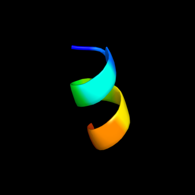
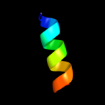
| 1 |
|
PDB 2bn8 chain A
Region: 15 - 23
Aligned: 9
Modelled: 9
Confidence: 8.5%
Identity: 33%
PDB header:cell cycle protein
Chain: A: PDB Molecule:cell division activator ceda;
PDBTitle: solution structure and interactions of the e.coli cell2 division activator protein ceda
Phyre2
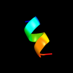
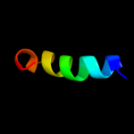
| 2 |
|
PDB 2d35 chain A
Region: 15 - 24
Aligned: 10
Modelled: 10
Confidence: 8.1%
Identity: 30%
PDB header:cell cycle
Chain: A: PDB Molecule:cell division activator ceda;
PDBTitle: solution structure of cell division reactivation factor,2 ceda
Phyre2
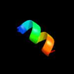
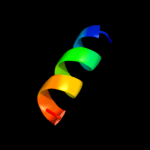
| 3 |
|
PDB 3bux chain B domain 1
Region: 12 - 37
Aligned: 18
Modelled: 26
Confidence: 7.4%
Identity: 33%
Fold: EF Hand-like
Superfamily: EF-hand
Family: EF-hand modules in multidomain proteins
Phyre2
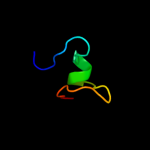
| 4 |
|
PDB 3dlq chain I domain 1
Region: 8 - 21
Aligned: 14
Modelled: 14
Confidence: 6.5%
Identity: 29%
Fold: 4-helical cytokines
Superfamily: 4-helical cytokines
Family: Interferons/interleukin-10 (IL-10)
Phyre2
| 5 |
|
PDB 2bid chain A
Region: 9 - 29
Aligned: 21
Modelled: 21
Confidence: 6.4%
Identity: 33%
Fold: Toxins' membrane translocation domains
Superfamily: Bcl-2 inhibitors of programmed cell death
Family: Bcl-2 inhibitors of programmed cell death
Phyre2
| 6 |
|
PDB 3arc chain L
Region: 32 - 44
Aligned: 13
Modelled: 13
Confidence: 5.7%
Identity: 46%
PDB header:electron transport, photosynthesis
Chain: L: PDB Molecule:photosystem ii reaction center protein l;
PDBTitle: crystal structure of oxygen-evolving photosystem ii at 1.9 angstrom2 resolution
Phyre2
|
| Detailed template information | |
Due to computational demand, binding site predictions are not run for batch jobs
If you want to predict binding sites, please manually submit your model of choice to 3DLigandSite
Phyre is for academic use only
| Please cite: Protein structure prediction on
the web: a case study using the Phyre server |
| Kelley LA and Sternberg MJE. Nature Protocols
4, 363 - 371 (2009) [pdf] [Import into BibTeX] |
| |
| If you use the binding site
predictions from 3DLigandSite, please also cite: |
| 3DLigandSite: predicting ligand-binding sites using similar structures. |
| Wass MN, Kelley LA and Sternberg
MJ Nucleic Acids Research 38, W469-73 (2010) [PubMed] |
| |
|
|
|
|