| 1 | c3c8fA_
|
|
 |
99.9 |
20 |
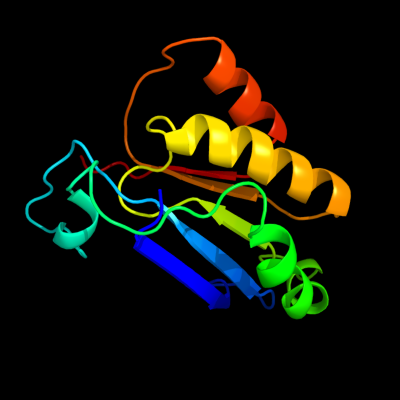
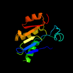
PDB header:oxidoreductase
Chain: A: PDB Molecule:pyruvate formate-lyase 1-activating enzyme;
PDBTitle: 4fe-4s-pyruvate formate-lyase activating enzyme with2 partially disordered adomet
|
| 2 | d1tv8a_
|
|
 |
99.5 |
19 |
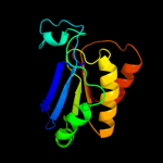
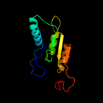
Fold:TIM beta/alpha-barrel
Superfamily:Radical SAM enzymes
Family:MoCo biosynthesis proteins |
| 3 | c2yx0A_
|
|
 |
99.5 |
12 |
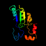
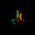
PDB header:metal binding protein
Chain: A: PDB Molecule:radical sam enzyme;
PDBTitle: crystal structure of p. horikoshii tyw1
|
| 4 | c3rfaA_
|
|
 |
99.1 |
14 |
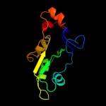
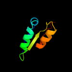
PDB header:oxidoreductase
Chain: A: PDB Molecule:ribosomal rna large subunit methyltransferase n;
PDBTitle: x-ray structure of rlmn from escherichia coli in complex with s-2 adenosylmethionine
|
| 5 | c2z2uA_
|
|
 |
98.8 |
10 |
PDB header:metal binding protein
Chain: A: PDB Molecule:upf0026 protein mj0257;
PDBTitle: crystal structure of archaeal tyw1
|
| 6 | c2a5hC_
|
|
 |
98.8 |
14 |
PDB header:isomerase
Chain: C: PDB Molecule:l-lysine 2,3-aminomutase;
PDBTitle: 2.1 angstrom x-ray crystal structure of lysine-2,3-aminomutase from2 clostridium subterminale sb4, with michaelis analog (l-alpha-lysine3 external aldimine form of pyridoxal-5'-phosphate).
|
| 7 | c3canA_
|
|
 |
98.5 |
15 |
PDB header:lyase activator
Chain: A: PDB Molecule:pyruvate-formate lyase-activating enzyme;
PDBTitle: crystal structure of a domain of pyruvate-formate lyase-activating2 enzyme from bacteroides vulgatus atcc 8482
|
| 8 | d1r30a_
|
|
 |
98.1 |
11 |
Fold:TIM beta/alpha-barrel
Superfamily:Radical SAM enzymes
Family:Biotin synthase |
| 9 | c1r30A_
|
|
 |
98.1 |
11 |
PDB header:transferase
Chain: A: PDB Molecule:biotin synthase;
PDBTitle: the crystal structure of biotin synthase, an s-2 adenosylmethionine-dependent radical enzyme
|
| 10 | c3t7vA_
|
|
 |
98.0 |
16 |
PDB header:transferase
Chain: A: PDB Molecule:methylornithine synthase pylb;
PDBTitle: crystal structure of methylornithine synthase (pylb)
|
| 11 | c3cixA_
|
|
 |
97.9 |
15 |
PDB header:adomet binding protein
Chain: A: PDB Molecule:fefe-hydrogenase maturase;
PDBTitle: x-ray structure of the [fefe]-hydrogenase maturase hyde from2 thermotoga maritima in complex with thiocyanate
|
| 12 | d1olta_
|
|
 |
97.2 |
15 |
Fold:TIM beta/alpha-barrel
Superfamily:Radical SAM enzymes
Family:Oxygen-independent coproporphyrinogen III oxidase HemN |
| 13 | d1lbua2
|
|
 |
33.4 |
27 |
Fold:Hedgehog/DD-peptidase
Superfamily:Hedgehog/DD-peptidase
Family:Muramoyl-pentapeptide carboxypeptidase |
| 14 | c2klxA_
|
|
 |
30.9 |
41 |
PDB header:oxidoreductase
Chain: A: PDB Molecule:glutaredoxin;
PDBTitle: solution structure of glutaredoxin from bartonella henselae str.2 houston
|
| 15 | c2hl7A_
|
|
 |
28.1 |
28 |
PDB header:oxidoreductase
Chain: A: PDB Molecule:cytochrome c-type biogenesis protein ccmh;
PDBTitle: crystal structure of the periplasmic domain of ccmh from pseudomonas2 aeruginosa
|
| 16 | c2qgqF_
|
|
 |
27.8 |
17 |
PDB header:structural genomics, unknown function
Chain: F: PDB Molecule:protein tm_1862;
PDBTitle: crystal structure of tm_1862 from thermotoga maritima.2 northeast structural genomics consortium target vr77
|
| 17 | c2kw0A_
|
|
 |
26.8 |
18 |
PDB header:oxidoreductase
Chain: A: PDB Molecule:ccmh protein;
PDBTitle: solution structure of n-terminal domain of ccmh from escherichia.coli
|
| 18 | c2kl5A_
|
|
 |
25.7 |
29 |
PDB header:structural genomics, unknown function
Chain: A: PDB Molecule:uncharacterized protein yutd;
PDBTitle: solution nmr structure of protein yutd from b.subtilis, northeast2 structural genomics consortium target sr232
|
| 19 | c3gv1A_
|
|
 |
25.2 |
30 |
PDB header:structural genomics, unknown function
Chain: A: PDB Molecule:disulfide interchange protein;
PDBTitle: crystal structure of disulfide interchange protein from neisseria2 gonorrhoeae
|
| 20 | c3kc2A_
|
|
 |
24.0 |
10 |
PDB header:hydrolase
Chain: A: PDB Molecule:uncharacterized protein ykr070w;
PDBTitle: crystal structure of mitochondrial had-like phosphatase from2 saccharomyces cerevisiae
|
| 21 | c3gn3B_ |
|
not modelled |
21.3 |
23 |
PDB header:structural genomics, unknown function
Chain: B: PDB Molecule:putative protein-disulfide isomerase;
PDBTitle: crystal structure of a putative protein-disulfide isomerase from2 pseudomonas syringae to 2.5a resolution.
|
| 22 | c3h93A_ |
|
not modelled |
19.0 |
22 |
PDB header:transcription regulator
Chain: A: PDB Molecule:thiol:disulfide interchange protein dsba;
PDBTitle: crystal structure of pseudomonas aeruginosa dsba
|
| 23 | c3dvwA_ |
|
not modelled |
17.6 |
22 |
PDB header:oxidoreductase
Chain: A: PDB Molecule:thiol:disulfide interchange protein dsba;
PDBTitle: crystal structure of reduced dsba1 from neisseria2 meningitidis
|
| 24 | c2k0rA_ |
|
not modelled |
16.9 |
19 |
PDB header:oxidoreductase
Chain: A: PDB Molecule:thiol:disulfide interchange protein dsbd;
PDBTitle: solution structure of the c103s mutant of the n-terminal2 domain of dsbd from neisseria meningitidis
|
| 25 | c1v57A_ |
|
not modelled |
16.6 |
24 |
PDB header:isomerase
Chain: A: PDB Molecule:thiol:disulfide interchange protein dsbg;
PDBTitle: crystal structure of the disulfide bond isomerase dsbg
|
| 26 | c2k9hA_ |
|
not modelled |
16.5 |
38 |
PDB header:metal binding protein
Chain: A: PDB Molecule:glycoprotein;
PDBTitle: the hantavirus glycoprotein g1 tail contains a dual cchc-2 type classical zinc fingers
|
| 27 | c3l9vE_ |
|
not modelled |
16.4 |
31 |
PDB header:oxidoreductase
Chain: E: PDB Molecule:putative thiol-disulfide isomerase or thioredoxin;
PDBTitle: crystal structure of salmonella enterica serovar typhimurium srga
|
| 28 | d2atcb2 |
|
not modelled |
16.2 |
33 |
Fold:Rubredoxin-like
Superfamily:Aspartate carbamoyltransferase, Regulatory-chain, C-terminal domain
Family:Aspartate carbamoyltransferase, Regulatory-chain, C-terminal domain |
| 29 | c2l7xA_ |
|
not modelled |
16.1 |
40 |
PDB header:viral protein
Chain: A: PDB Molecule:envelope glycoprotein;
PDBTitle: crimean congo hemorrhagic fever gn zinc finger
|
| 30 | c3ct7E_ |
|
not modelled |
16.0 |
15 |
PDB header:isomerase
Chain: E: PDB Molecule:d-allulose-6-phosphate 3-epimerase;
PDBTitle: crystal structure of d-allulose 6-phosphate 3-epimerase2 from escherichia coli k-12
|
| 31 | c3c7mB_ |
|
not modelled |
15.5 |
23 |
PDB header:oxidoreductase
Chain: B: PDB Molecule:thiol:disulfide interchange protein dsba-like;
PDBTitle: crystal structure of reduced dsbl
|
| 32 | c3feuA_ |
|
not modelled |
14.3 |
22 |
PDB header:oxidoreductase
Chain: A: PDB Molecule:putative lipoprotein;
PDBTitle: crystal structure of dsba-like thioredoxin domain vf_a0457 from vibrio2 fischeri
|
| 33 | c1nl0G_ |
|
not modelled |
14.1 |
50 |
PDB header:immune system
Chain: G: PDB Molecule:factor ix;
PDBTitle: crystal structure of human factor ix gla domain in complex2 of an inhibitory antibody, 10c12
|
| 34 | c3cu2A_ |
|
not modelled |
14.0 |
11 |
PDB header:isomerase
Chain: A: PDB Molecule:ribulose-5-phosphate 3-epimerase;
PDBTitle: crystal structure of ribulose-5-phosphate 3-epimerase (yp_718263.1)2 from haemophilus somnus 129pt at 1.91 a resolution
|
| 35 | d1vsra_ |
|
not modelled |
13.4 |
42 |
Fold:Restriction endonuclease-like
Superfamily:Restriction endonuclease-like
Family:Very short patch repair (VSR) endonuclease |
| 36 | d1beda_ |
|
not modelled |
13.3 |
22 |
Fold:Thioredoxin fold
Superfamily:Thioredoxin-like
Family:DsbA-like |
| 37 | d1ckqa_ |
|
not modelled |
13.2 |
22 |
Fold:Restriction endonuclease-like
Superfamily:Restriction endonuclease-like
Family:Restriction endonuclease EcoRI |
| 38 | d2fzcb2 |
|
not modelled |
13.2 |
33 |
Fold:Rubredoxin-like
Superfamily:Aspartate carbamoyltransferase, Regulatory-chain, C-terminal domain
Family:Aspartate carbamoyltransferase, Regulatory-chain, C-terminal domain |
| 39 | d1abaa_ |
|
not modelled |
13.1 |
20 |
Fold:Thioredoxin fold
Superfamily:Thioredoxin-like
Family:Thioltransferase |
| 40 | d1eysc_ |
|
not modelled |
13.0 |
22 |
Fold:Multiheme cytochromes
Superfamily:Multiheme cytochromes
Family:Photosynthetic reaction centre (cytochrome subunit) |
| 41 | c1eysC_ |
|
not modelled |
13.0 |
22 |
PDB header:electron transport
Chain: C: PDB Molecule:photosynthetic reaction center;
PDBTitle: crystal structure of photosynthetic reaction center from a2 thermophilic bacterium, thermochromatium tepidum
|
| 42 | c2znmA_ |
|
not modelled |
12.6 |
19 |
PDB header:oxidoreductase
Chain: A: PDB Molecule:thiol:disulfide interchange protein dsba;
PDBTitle: oxidoreductase nmdsba3 from neisseria meningitidis
|
| 43 | c2khpA_ |
|
not modelled |
12.4 |
35 |
PDB header:electron transport
Chain: A: PDB Molecule:glutaredoxin;
PDBTitle: solution structure of glutaredoxin from brucella melitensis
|
| 44 | d1egoa_ |
|
not modelled |
12.3 |
31 |
Fold:Thioredoxin fold
Superfamily:Thioredoxin-like
Family:Thioltransferase |
| 45 | c2bpbB_ |
|
not modelled |
12.3 |
12 |
PDB header:oxidoreductase
Chain: B: PDB Molecule:sulfite\:cytochrome c oxidoreductase subunit b;
PDBTitle: sulfite dehydrogenase from starkeya novella
|
| 46 | c2he3A_ |
|
not modelled |
12.1 |
16 |
PDB header:oxidoreductase
Chain: A: PDB Molecule:glutathione peroxidase 2;
PDBTitle: crystal structure of the selenocysteine to cysteine mutant of human2 glutathionine peroxidase 2 (gpx2)
|
| 47 | d1pg5b2 |
|
not modelled |
12.0 |
33 |
Fold:Rubredoxin-like
Superfamily:Aspartate carbamoyltransferase, Regulatory-chain, C-terminal domain
Family:Aspartate carbamoyltransferase, Regulatory-chain, C-terminal domain |
| 48 | d2i5nc1 |
|
not modelled |
11.4 |
12 |
Fold:Multiheme cytochromes
Superfamily:Multiheme cytochromes
Family:Photosynthetic reaction centre (cytochrome subunit) |
| 49 | c2jblC_ |
|
not modelled |
11.4 |
12 |
PDB header:electron transport
Chain: C: PDB Molecule:photosynthetic reaction center cytochrome c
PDBTitle: photosynthetic reaction center from blastochloris viridis
|
| 50 | d1wjpa2 |
|
not modelled |
11.1 |
30 |
Fold:beta-beta-alpha zinc fingers
Superfamily:beta-beta-alpha zinc fingers
Family:Classic zinc finger, C2H2 |
| 51 | c2be7E_ |
|
not modelled |
10.5 |
33 |
PDB header:transferase
Chain: E: PDB Molecule:aspartate carbamoyltransferase regulatory chain;
PDBTitle: crystal structure of the unliganded (t-state) aspartate2 transcarbamoylase of the psychrophilic bacterium moritella profunda
|
| 52 | d1t3ba1 |
|
not modelled |
9.3 |
29 |
Fold:Thioredoxin fold
Superfamily:Thioredoxin-like
Family:DsbC/DsbG C-terminal domain-like |
| 53 | d1h75a_ |
|
not modelled |
9.2 |
14 |
Fold:Thioredoxin fold
Superfamily:Thioredoxin-like
Family:Thioltransferase |
| 54 | d3c7bb3 |
|
not modelled |
9.2 |
20 |
Fold:Nitrite and sulphite reductase 4Fe-4S domain-like
Superfamily:Nitrite and sulphite reductase 4Fe-4S domain-like
Family:Nitrite and sulphite reductase 4Fe-4S domain-like |
| 55 | c2e2zA_ |
|
not modelled |
9.0 |
13 |
PDB header:protein transport, chaperone regulator
Chain: A: PDB Molecule:tim15;
PDBTitle: solution nmr structure of yeast tim15, co-chaperone of2 mitochondrial hsp70
|
| 56 | d1x5wa2 |
|
not modelled |
8.9 |
29 |
Fold:beta-beta-alpha zinc fingers
Superfamily:beta-beta-alpha zinc fingers
Family:Classic zinc finger, C2H2 |
| 57 | c3gr7A_ |
|
not modelled |
8.5 |
11 |
PDB header:oxidoreductase
Chain: A: PDB Molecule:nadph dehydrogenase;
PDBTitle: structure of oye from geobacillus kaustophilus, hexagonal2 crystal form
|
| 58 | d1atxa_ |
|
not modelled |
8.5 |
39 |
Fold:Defensin-like
Superfamily:Defensin-like
Family:Defensin |
| 59 | c3kwpA_ |
|
not modelled |
8.2 |
15 |
PDB header:transferase
Chain: A: PDB Molecule:predicted methyltransferase;
PDBTitle: crystal structure of putative methyltransferase from lactobacillus2 brevis
|
| 60 | d2hfqa1 |
|
not modelled |
8.1 |
43 |
Fold:NE1680-like
Superfamily:NE1680-like
Family:NE1680-like |
| 61 | c2hfqA_ |
|
not modelled |
8.1 |
43 |
PDB header:structural genomics, unknown function
Chain: A: PDB Molecule:hypothetical protein;
PDBTitle: nmr structure of protein ne1680 from nitrosomonas europaea:2 northeast structural genomics consortium target net5
|
| 62 | d1pbya1 |
|
not modelled |
8.1 |
17 |
Fold:Cytochrome c
Superfamily:Cytochrome c
Family:Quinohemoprotein amine dehydrogenase A chain, domains 1 and 2 |
| 63 | d1eeja1 |
|
not modelled |
8.0 |
25 |
Fold:Thioredoxin fold
Superfamily:Thioredoxin-like
Family:DsbC/DsbG C-terminal domain-like |
| 64 | c2elpA_ |
|
not modelled |
8.0 |
50 |
PDB header:transcription
Chain: A: PDB Molecule:zinc finger protein 406;
PDBTitle: solution structure of the 13th c2h2 zinc finger of human2 zinc finger protein 406
|
| 65 | d2akja3 |
|
not modelled |
7.9 |
25 |
Fold:Nitrite and sulphite reductase 4Fe-4S domain-like
Superfamily:Nitrite and sulphite reductase 4Fe-4S domain-like
Family:Nitrite and sulphite reductase 4Fe-4S domain-like |
| 66 | c2remB_ |
|
not modelled |
7.8 |
21 |
PDB header:oxidoreductase
Chain: B: PDB Molecule:disulfide oxidoreductase;
PDBTitle: crystal structure of oxidoreductase dsba from xylella2 fastidiosa
|
| 67 | d1cw0a_ |
|
not modelled |
7.8 |
42 |
Fold:Restriction endonuclease-like
Superfamily:Restriction endonuclease-like
Family:Very short patch repair (VSR) endonuclease |
| 68 | c3bciA_ |
|
not modelled |
7.6 |
33 |
PDB header:oxidoreductase
Chain: A: PDB Molecule:disulfide bond protein a;
PDBTitle: crystal structure of staphylococcus aureus dsba
|
| 69 | d1z6ma1 |
|
not modelled |
7.6 |
38 |
Fold:Thioredoxin fold
Superfamily:Thioredoxin-like
Family:DsbA-like |
| 70 | c3omzG_ |
|
not modelled |
7.5 |
25 |
PDB header:immune system
Chain: G: PDB Molecule:human vdelta1 gamma delta t cell receptor delta1a/b-3;
PDBTitle: crystal structure of mica-specific human gamma delta t cell receptor
|
| 71 | c3hf3A_ |
|
not modelled |
7.3 |
11 |
PDB header:oxidoreductase
Chain: A: PDB Molecule:chromate reductase;
PDBTitle: old yellow enzyme from thermus scotoductus sa-01
|
| 72 | d1uxja1 |
|
not modelled |
7.3 |
18 |
Fold:NAD(P)-binding Rossmann-fold domains
Superfamily:NAD(P)-binding Rossmann-fold domains
Family:LDH N-terminal domain-like |
| 73 | d1r7ha_ |
|
not modelled |
7.2 |
13 |
Fold:Thioredoxin fold
Superfamily:Thioredoxin-like
Family:Thioltransferase |
| 74 | c1u83A_ |
|
not modelled |
7.2 |
15 |
PDB header:lyase
Chain: A: PDB Molecule:phosphosulfolactate synthase;
PDBTitle: psl synthase from bacillus subtilis
|
| 75 | d1u83a_ |
|
not modelled |
7.2 |
15 |
Fold:TIM beta/alpha-barrel
Superfamily:(2r)-phospho-3-sulfolactate synthase ComA
Family:(2r)-phospho-3-sulfolactate synthase ComA |
| 76 | d2ouwa1 |
|
not modelled |
7.1 |
17 |
Fold:AhpD-like
Superfamily:AhpD-like
Family:TTHA0727-like |
| 77 | c1i7mD_ |
|
not modelled |
7.0 |
18 |
PDB header:lyase
Chain: D: PDB Molecule:s-adenosylmethionine decarboxylase beta chain;
PDBTitle: human s-adenosylmethionine decarboxylase with covalently bound2 pyruvoyl group and complexed with 4-amidinoindan-1-one-2'-3 amidinohydrazone
|
| 78 | d1vkea_ |
|
not modelled |
7.0 |
11 |
Fold:AhpD-like
Superfamily:AhpD-like
Family:CMD-like |
| 79 | c2w95B_ |
|
not modelled |
6.9 |
11 |
PDB header:cell adhesion
Chain: B: PDB Molecule:discoidin-1 subunit a;
PDBTitle: structure of the discoidin i from dictyostelium discoideum2 in complex with galnac at 1.75 angstrom resolution
|
| 80 | d1fova_ |
|
not modelled |
6.9 |
23 |
Fold:Thioredoxin fold
Superfamily:Thioredoxin-like
Family:Thioltransferase |
| 81 | d1jmxa1 |
|
not modelled |
6.7 |
19 |
Fold:Cytochrome c
Superfamily:Cytochrome c
Family:Quinohemoprotein amine dehydrogenase A chain, domains 1 and 2 |
| 82 | d1aopa4 |
|
not modelled |
6.5 |
40 |
Fold:Nitrite and sulphite reductase 4Fe-4S domain-like
Superfamily:Nitrite and sulphite reductase 4Fe-4S domain-like
Family:Nitrite and sulphite reductase 4Fe-4S domain-like |
| 83 | d1un2a_ |
|
not modelled |
6.5 |
36 |
Fold:Thioredoxin fold
Superfamily:Thioredoxin-like
Family:DsbA-like |
| 84 | d1fvka_ |
|
not modelled |
6.4 |
36 |
Fold:Thioredoxin fold
Superfamily:Thioredoxin-like
Family:DsbA-like |
| 85 | d1dl6a_ |
|
not modelled |
6.3 |
15 |
Fold:Rubredoxin-like
Superfamily:Zinc beta-ribbon
Family:Transcriptional factor domain |
| 86 | d1tzaa_ |
|
not modelled |
6.3 |
39 |
Fold:Immunoglobulin-like beta-sandwich
Superfamily:ApaG-like
Family:ApaG-like |
| 87 | d2v4jb3 |
|
not modelled |
6.2 |
18 |
Fold:Nitrite and sulphite reductase 4Fe-4S domain-like
Superfamily:Nitrite and sulphite reductase 4Fe-4S domain-like
Family:Nitrite and sulphite reductase 4Fe-4S domain-like |
| 88 | c1t3bA_ |
|
not modelled |
6.2 |
29 |
PDB header:isomerase
Chain: A: PDB Molecule:thiol:disulfide interchange protein dsbc;
PDBTitle: x-ray structure of dsbc from haemophilus influenzae
|
| 89 | d1adta2 |
|
not modelled |
6.1 |
23 |
Fold:Zn-binding domains of ADDBP
Superfamily:Zn-binding domains of ADDBP
Family:Zn-binding domains of ADDBP |
| 90 | c2f1eA_ |
|
not modelled |
6.1 |
26 |
PDB header:structural genomics, unknown function
Chain: A: PDB Molecule:protein apag;
PDBTitle: solution structure of apag protein
|
| 91 | d1ekqa_ |
|
not modelled |
6.1 |
13 |
Fold:Ribokinase-like
Superfamily:Ribokinase-like
Family:Thiamin biosynthesis kinases |
| 92 | d2o4da1 |
|
not modelled |
6.0 |
15 |
Fold:AhpD-like
Superfamily:AhpD-like
Family:Atu0492-like |
| 93 | d1r4wa_ |
|
not modelled |
6.0 |
21 |
Fold:Thioredoxin fold
Superfamily:Thioredoxin-like
Family:DsbA-like |
| 94 | d2drpa2 |
|
not modelled |
5.9 |
60 |
Fold:beta-beta-alpha zinc fingers
Superfamily:beta-beta-alpha zinc fingers
Family:Classic zinc finger, C2H2 |
| 95 | c2k5cA_ |
|
not modelled |
5.8 |
17 |
PDB header:metal binding protein
Chain: A: PDB Molecule:uncharacterized protein pf0385;
PDBTitle: nmr structure for pf0385
|
| 96 | d2fug21 |
|
not modelled |
5.8 |
12 |
Fold:Thioredoxin fold
Superfamily:Thioredoxin-like
Family:NQO2-like |
| 97 | d1twia2 |
|
not modelled |
5.7 |
14 |
Fold:TIM beta/alpha-barrel
Superfamily:PLP-binding barrel
Family:Alanine racemase-like, N-terminal domain |
| 98 | c3f4tA_ |
|
not modelled |
5.7 |
17 |
PDB header:oxidoreductase
Chain: A: PDB Molecule:putative uncharacterized protein;
PDBTitle: crystal structure of wolbachia pipientis alpha-dsba1 c97a/c146a
|
| 99 | d1tdha3 |
|
not modelled |
5.6 |
29 |
Fold:Glucocorticoid receptor-like (DNA-binding domain)
Superfamily:Glucocorticoid receptor-like (DNA-binding domain)
Family:C-terminal, Zn-finger domain of MutM-like DNA repair proteins |