Phyre Home Retrieve Phyre Job Id
Emaill.a.kelley@imperial.ac.uk
DescriptionP0ADZ4
DateThu Jan 5 11:22:08 GMT 2012
Unique Job ID61c270b61956df4f

Summary 

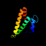
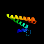
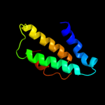
Top model
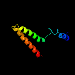
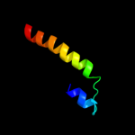
Image coloured by rainbow N → C terminus
Model (left) based on template d1vs5o1
Top template information
Fold:S15/NS1 RNA-binding domain
Superfamily:S15/NS1 RNA-binding domain
Family:Ribosomal protein S15
Confidence and coverage
Confidence:100.0% Coverage: 99%
88 residues ( 99% of your sequence) have been modelled with 100.0% confidence by the single highest scoring template.
3D viewing
Interactive 3D view in Jmol

Sequence analysis 

Secondary structure and disorder prediction 

   1........10.........20.........30.........40.........50.........60
Sequence  MSLSTEATAKIVSEFGRDANDTGSTEVQVALLTAQINHLQGHFAEHKKDHHSRRGLLRMV
Secondary structure 


SS confidence 


Disorder  ????


????


??


Disorder confidence 


   .........70.........80.........
Sequence  SQRRKLLDYLKRKDVARYTQLIERLGLRR
Secondary structure 


SS confidence 


Disorder 


????
Disorder confidence 


Confidence Key
High(9)                    Low (0)
?Disordered
Alpha helix
Beta strand

Domain analysis 

Hover over an aligned region to see model and summary info

Please note, only up to the top 20 hits are modelled to reduce computer load

RankAligned region

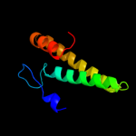
PDB 1vs5 chain O domain 1

3D model

Region: 2 - 89
Aligned: 88
Modelled: 88
Confidence: 100.0%
Identity: 99%
Fold: S15/NS1 RNA-binding domain
Superfamily: S15/NS1 RNA-binding domain
Family: Ribosomal protein S15

Phyre2

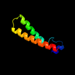
PDB 1g1x chain B

3D model

Region: 2 - 88
Aligned: 87
Modelled: 87
Confidence: 100.0%
Identity: 59%
Fold: S15/NS1 RNA-binding domain
Superfamily: S15/NS1 RNA-binding domain
Family: Ribosomal protein S15

Phyre2

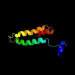
PDB 1a32 chain A

3D model

Region: 3 - 87
Aligned: 85
Modelled: 85
Confidence: 100.0%
Identity: 59%
Fold: S15/NS1 RNA-binding domain
Superfamily: S15/NS1 RNA-binding domain
Family: Ribosomal protein S15

Phyre2

PDB 3ulw chain A

3D model

Region: 3 - 87
Aligned: 85
Modelled: 85
Confidence: 100.0%
Identity: 54%
PDB header:ribosomal protein
Chain: A: PDB Molecule:30s ribosomal protein s15;
PDBTitle: 30s ribosomal protein s15 from campylobacter jejuni

Phyre2

PDB 1kuq chain A

3D model

Region: 5 - 87
Aligned: 83
Modelled: 83
Confidence: 100.0%
Identity: 61%
Fold: S15/NS1 RNA-binding domain
Superfamily: S15/NS1 RNA-binding domain
Family: Ribosomal protein S15

Phyre2

PDB 3bbn chain O

3D model

Region: 7 - 89
Aligned: 83
Modelled: 83
Confidence: 100.0%
Identity: 35%
PDB header:ribosome
Chain: O: PDB Molecule:ribosomal protein s15;
PDBTitle: homology model for the spinach chloroplast 30s subunit2 fitted to 9.4a cryo-em map of the 70s chlororibosome.

Phyre2

PDB 2xzm chain O

3D model

Region: 12 - 74
Aligned: 63
Modelled: 63
Confidence: 99.4%
Identity: 16%
PDB header:ribosome
Chain: O: PDB Molecule:rps13e;
PDBTitle: crystal structure of the eukaryotic 40s ribosomal2 subunit in complex with initiation factor 1. this file3 contains the 40s subunit and initiation factor for4 molecule 1

Phyre2

PDB 3izb chain O

3D model

Region: 12 - 87
Aligned: 76
Modelled: 76
Confidence: 99.4%
Identity: 11%
PDB header:ribosome
Chain: O: PDB Molecule:40s ribosomal protein rps13 (s15p);
PDBTitle: localization of the small subunit ribosomal proteins into a 6.1 a2 cryo-em map of saccharomyces cerevisiae translating 80s ribosome

Phyre2

PDB 1ysh chain E

3D model

Region: 2 - 74
Aligned: 69
Modelled: 73
Confidence: 98.3%
Identity: 20%
PDB header:structural protein/rna
Chain: E: PDB Molecule:40s ribosomal protein s13;
PDBTitle: localization and dynamic behavior of ribosomal protein l30e

Phyre2

PDB 1s1h chain O

3D model

Region: 12 - 69
Aligned: 58
Modelled: 58
Confidence: 96.6%
Identity: 14%
PDB header:ribosome
Chain: O: PDB Molecule:40s ribosomal protein s13;
PDBTitle: structure of the ribosomal 80s-eef2-sordarin complex from2 yeast obtained by docking atomic models for rna and protein3 components into a 11.7 a cryo-em map. this file, 1s1h,4 contains 40s subunit. the 60s ribosomal subunit is in file5 1s1i.

Phyre2

PDB 1y0u chain A

3D model

Region: 63 - 87
Aligned: 23
Modelled: 25
Confidence: 32.2%
Identity: 30%
Fold: DNA/RNA-binding 3-helical bundle
Superfamily: "Winged helix" DNA-binding domain
Family: ArsR-like transcriptional regulators

Phyre2

PDB 2y50 chain A

3D model

Region: 64 - 86
Aligned: 23
Modelled: 23
Confidence: 25.8%
Identity: 26%
PDB header:hydrolase
Chain: A: PDB Molecule:collagenase;
PDBTitle: crystal structure of collagenase g from clostridium2 histolyticum at 2.80 angstrom resolution

Phyre2

PDB 2ke4 chain A

3D model

Region: 9 - 46
Aligned: 38
Modelled: 38
Confidence: 18.8%
Identity: 21%
PDB header:membrane protein
Chain: A: PDB Molecule:cdc42-interacting protein 4;
PDBTitle: the nmr structure of the tc10 and cdc42 interacting domain2 of cip4

Phyre2

PDB 2zvn chain F

3D model

Region: 27 - 71
Aligned: 45
Modelled: 45
Confidence: 17.0%
Identity: 16%
PDB header:signaling protein/transcription
Chain: F: PDB Molecule:nf-kappa-b essential modulator;
PDBTitle: nemo cozi domain incomplex with diubiquitin in p2121212 space group

Phyre2

PDB 3f1i chain H

3D model

Region: 28 - 86
Aligned: 59
Modelled: 59
Confidence: 12.4%
Identity: 17%
PDB header:protein binding
Chain: H: PDB Molecule:hepatocyte growth factor-regulated tyrosine kinase
PDBTitle: human escrt-0 core complex

Phyre2

PDB 2vog chain B

3D model

Region: 25 - 30
Aligned: 6
Modelled: 6
Confidence: 8.7%
Identity: 67%
PDB header:apoptosis
Chain: B: PDB Molecule:bcl-2-modifying factor;
PDBTitle: structure of mouse a1 bound to the bmf bh3-domain

Phyre2

PDB 2v4h chain A

3D model

Region: 28 - 71
Aligned: 44
Modelled: 44
Confidence: 8.1%
Identity: 16%
PDB header:transcription
Chain: A: PDB Molecule:nf-kappa-b essential modulator;
PDBTitle: nemo cc2-lz domain - 1d5 darpin complex

Phyre2

PDB 1sxj chain D domain 1

3D model

Region: 1 - 39
Aligned: 39
Modelled: 39
Confidence: 7.1%
Identity: 18%
Fold: post-AAA+ oligomerization domain-like
Superfamily: post-AAA+ oligomerization domain-like
Family: DNA polymerase III clamp loader subunits, C-terminal domain

Phyre2

PDB 2f5x chain C

3D model

Region: 72 - 89
Aligned: 18
Modelled: 18
Confidence: 6.7%
Identity: 17%
PDB header:transport protein
Chain: C: PDB Molecule:bugd;
PDBTitle: structure of periplasmic binding protein bugd

Phyre2

PDB 3nqw chain B

3D model

Region: 5 - 87
Aligned: 77
Modelled: 83
Confidence: 6.7%
Identity: 12%
PDB header:hydrolase
Chain: B: PDB Molecule:cg11900;
PDBTitle: a metazoan ortholog of spot hydrolyzes ppgpp and plays a role in2 starvation responses

Phyre2
1

d1vs5o1
2

d1g1xb_
3

d1a32a_
4

c3ulwA_
5

d1kuqa_
6

c3bbnO_
7

c2xzmO_
8

c3izbO_
9

c1yshE_
10

c1s1hO_
11

d1y0ua_
12

c2y50A_
13

c2ke4A_
14

c2zvnF_
15

c3f1iH_
16

c2vogB_
17

c2v4hA_
18

d1sxjd1
19

c2f5xC_
20

c3nqwB_
21


22


23


24


25


26


Detailed template information 

#
Template Alignment Coverage3D Model Confidence
% i.d. Template Information
1d1vs5o1


100.0 99 Fold:S15/NS1 RNA-binding domain
Superfamily:S15/NS1 RNA-binding domain
Family:Ribosomal protein S15
2d1g1xb_


100.0 59 Fold:S15/NS1 RNA-binding domain
Superfamily:S15/NS1 RNA-binding domain
Family:Ribosomal protein S15
3d1a32a_


100.0 59 Fold:S15/NS1 RNA-binding domain
Superfamily:S15/NS1 RNA-binding domain
Family:Ribosomal protein S15
4c3ulwA_


100.0 54 PDB header:ribosomal protein
Chain: A: PDB Molecule:30s ribosomal protein s15;
PDBTitle: 30s ribosomal protein s15 from campylobacter jejuni
5d1kuqa_


100.0 61 Fold:S15/NS1 RNA-binding domain
Superfamily:S15/NS1 RNA-binding domain
Family:Ribosomal protein S15
6c3bbnO_


100.0 35 PDB header:ribosome
Chain: O: PDB Molecule:ribosomal protein s15;
PDBTitle: homology model for the spinach chloroplast 30s subunit2 fitted to 9.4a cryo-em map of the 70s chlororibosome.
7c2xzmO_


99.4 16 PDB header:ribosome
Chain: O: PDB Molecule:rps13e;
PDBTitle: crystal structure of the eukaryotic 40s ribosomal2 subunit in complex with initiation factor 1. this file3 contains the 40s subunit and initiation factor for4 molecule 1
8c3izbO_


99.4 11 PDB header:ribosome
Chain: O: PDB Molecule:40s ribosomal protein rps13 (s15p);
PDBTitle: localization of the small subunit ribosomal proteins into a 6.1 a2 cryo-em map of saccharomyces cerevisiae translating 80s ribosome
9c1yshE_


98.3 20 PDB header:structural protein/rna
Chain: E: PDB Molecule:40s ribosomal protein s13;
PDBTitle: localization and dynamic behavior of ribosomal protein l30e
10c1s1hO_


96.6 14 PDB header:ribosome
Chain: O: PDB Molecule:40s ribosomal protein s13;
PDBTitle: structure of the ribosomal 80s-eef2-sordarin complex from2 yeast obtained by docking atomic models for rna and protein3 components into a 11.7 a cryo-em map. this file, 1s1h,4 contains 40s subunit. the 60s ribosomal subunit is in file5 1s1i.
11d1y0ua_


32.2 30 Fold:DNA/RNA-binding 3-helical bundle
Superfamily:"Winged helix" DNA-binding domain
Family:ArsR-like transcriptional regulators
12c2y50A_


25.8 26 PDB header:hydrolase
Chain: A: PDB Molecule:collagenase;
PDBTitle: crystal structure of collagenase g from clostridium2 histolyticum at 2.80 angstrom resolution
13c2ke4A_


18.8 21 PDB header:membrane protein
Chain: A: PDB Molecule:cdc42-interacting protein 4;
PDBTitle: the nmr structure of the tc10 and cdc42 interacting domain2 of cip4
14c2zvnF_


17.0 16 PDB header:signaling protein/transcription
Chain: F: PDB Molecule:nf-kappa-b essential modulator;
PDBTitle: nemo cozi domain incomplex with diubiquitin in p2121212 space group
15c3f1iH_


12.4 17 PDB header:protein binding
Chain: H: PDB Molecule:hepatocyte growth factor-regulated tyrosine kinase
PDBTitle: human escrt-0 core complex
16c2vogB_


8.7 67 PDB header:apoptosis
Chain: B: PDB Molecule:bcl-2-modifying factor;
PDBTitle: structure of mouse a1 bound to the bmf bh3-domain
17c2v4hA_


8.1 16 PDB header:transcription
Chain: A: PDB Molecule:nf-kappa-b essential modulator;
PDBTitle: nemo cc2-lz domain - 1d5 darpin complex
18d1sxjd1


7.1 18 Fold:post-AAA+ oligomerization domain-like
Superfamily:post-AAA+ oligomerization domain-like
Family:DNA polymerase III clamp loader subunits, C-terminal domain
19c2f5xC_


6.7 17 PDB header:transport protein
Chain: C: PDB Molecule:bugd;
PDBTitle: structure of periplasmic binding protein bugd
20c3nqwB_


6.7 12 PDB header:hydrolase
Chain: B: PDB Molecule:cg11900;
PDBTitle: a metazoan ortholog of spot hydrolyzes ppgpp and plays a role in2 starvation responses
21c2pnvA_


not modelled 5.5 18 PDB header:membrane protein
Chain: A: PDB Molecule:small conductance calcium-activated potassium
PDBTitle: crystal structure of the leucine zipper domain of small-2 conductance ca2+-activated k+ (skca) channel from rattus3 norvegicus
22c3cuoB_


not modelled 5.5 24 PDB header:transcription regulator
Chain: B: PDB Molecule:uncharacterized hth-type transcriptional regulator ygav;
PDBTitle: crystal structure of the predicted dna-binding transcriptional2 regulator from e. coli
23c1m7lA_


not modelled 5.5 43 PDB header:sugar binding protein
Chain: A: PDB Molecule:pulmonary surfactant-associated protein d;
PDBTitle: solution structure of the coiled-coil trimerization domain2 from lung surfactant protein d
24d3crda_


not modelled 5.3 20 Fold:DEATH domain
Superfamily:DEATH domain
Family:Caspase recruitment domain, CARD
25d1biaa1


not modelled 5.3 30 Fold:DNA/RNA-binding 3-helical bundle
Superfamily:"Winged helix" DNA-binding domain
Family:Biotin repressor-like
26c2bmmA_


not modelled 5.2 6 PDB header:oxygen storage/transport
Chain: A: PDB Molecule:thermostable hemoglobin from thermobifida fusca;
PDBTitle: x-ray structure of a novel thermostable hemoglobin from the2 actinobacterium thermobifida fusca

Binding site prediction 

Due to computational demand, binding site predictions are not run for batch jobs

If you want to predict binding sites, please manually submit your model of choice to 3DLigandSite


Phyre is for academic use only

Please cite: Protein structure prediction on the web: a case study using the Phyre server
Kelley LA and Sternberg MJE. Nature Protocols 4, 363 - 371 (2009) [pdf] [Import into BibTeX]
 
If you use the binding site predictions from 3DLigandSite, please also cite:
3DLigandSite: predicting ligand-binding sites using similar structures.
Wass MN, Kelley LA and Sternberg MJ Nucleic Acids Research 38, W469-73 (2010) [PubMed]
 
© Structural Bioinformatics Group
Imperial College London
Lawrence Kelley, Benjamin Jefferys
Disclaimer
Terms and Conditions
Component software
Template detection: HHpred 1.51
Secondary structure prediction: Psi-pred 2.5
Disorder prediction: Disopred 2.4
Transmembrane prediction: Memsat_SVM
Multi-template modelling and ab initio: Poing 1.0