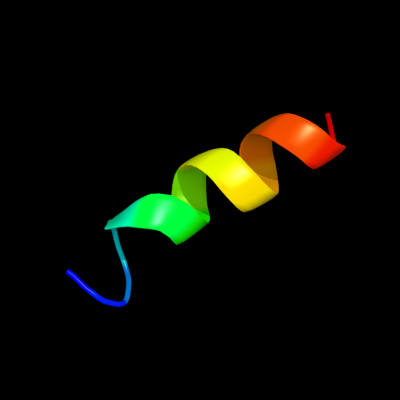
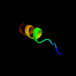
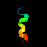
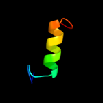
| 1 |
|
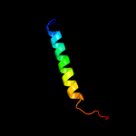
PDB 3p2o chain A
Region: 251 - 265
Aligned: 15
Modelled: 15
Confidence: 36.3%
Identity: 20%
PDB header:oxidoreductase, hydrolase
Chain: A: PDB Molecule:bifunctional protein fold;
PDBTitle: crystal structure of fold bifunctional protein from campylobacter2 jejuni
Phyre2
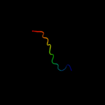
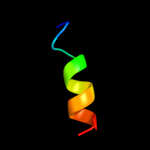
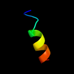
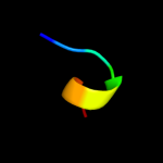
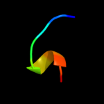
| 2 |
|
PDB 3l07 chain B
Region: 251 - 265
Aligned: 15
Modelled: 15
Confidence: 35.7%
Identity: 20%
PDB header:oxidoreductase,hydrolase
Chain: B: PDB Molecule:bifunctional protein fold;
PDBTitle: methylenetetrahydrofolate dehydrogenase/methenyltetrahydrofolate2 cyclohydrolase, putative bifunctional protein fold from francisella3 tularensis.
Phyre2
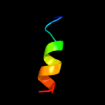
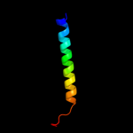
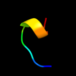
| 3 |
|
PDB 2c2x chain B
Region: 251 - 265
Aligned: 15
Modelled: 15
Confidence: 35.2%
Identity: 33%
PDB header:oxidoreductase
Chain: B: PDB Molecule:methylenetetrahydrofolate dehydrogenase-
PDBTitle: three dimensional structure of bifunctional2 methylenetetrahydrofolate dehydrogenase-cyclohydrolase3 from mycobacterium tuberculosis
Phyre2
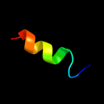
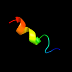
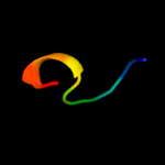
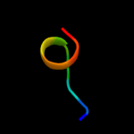
| 4 |
|
PDB 3p2o chain B
Region: 251 - 265
Aligned: 15
Modelled: 3
Confidence: 35.2%
Identity: 20%
PDB header:oxidoreductase, hydrolase
Chain: B: PDB Molecule:bifunctional protein fold;
PDBTitle: crystal structure of fold bifunctional protein from campylobacter2 jejuni
Phyre2
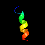
| 5 |
|
PDB 4a26 chain B
Region: 251 - 265
Aligned: 15
Modelled: 15
Confidence: 34.4%
Identity: 20%
PDB header:oxidoreductase
Chain: B: PDB Molecule:putative c-1-tetrahydrofolate synthase, cytoplasmic;
PDBTitle: the crystal structure of leishmania major n5,n10-2 methylenetetrahydrofolate dehydrogenase/cyclohydrolase
Phyre2
| 6 |
|
PDB 4a5o chain B
Region: 251 - 265
Aligned: 15
Modelled: 15
Confidence: 34.2%
Identity: 20%
PDB header:oxidoreductase
Chain: B: PDB Molecule:bifunctional protein fold;
PDBTitle: crystal structure of pseudomonas aeruginosa n5, n10-2 methylenetetrahydrofolate dehydrogenase-cyclohydrolase (fold)
Phyre2
| 7 |
|
PDB 1b0a chain A
Region: 251 - 265
Aligned: 15
Modelled: 15
Confidence: 34.0%
Identity: 20%
PDB header:oxidoreductase,hydrolase
Chain: A: PDB Molecule:protein (fold bifunctional protein);
PDBTitle: 5,10, methylene-tetrahydropholate2 dehydrogenase/cyclohydrolase from e coli.
Phyre2
| 8 |
|
PDB 1b0a chain A domain 1
Region: 251 - 265
Aligned: 15
Modelled: 15
Confidence: 27.0%
Identity: 20%
Fold: NAD(P)-binding Rossmann-fold domains
Superfamily: NAD(P)-binding Rossmann-fold domains
Family: Aminoacid dehydrogenase-like, C-terminal domain
Phyre2
| 9 |
|
PDB 1a4i chain B
Region: 251 - 265
Aligned: 15
Modelled: 15
Confidence: 25.8%
Identity: 20%
PDB header:oxidoreductase
Chain: B: PDB Molecule:methylenetetrahydrofolate dehydrogenase /
PDBTitle: human tetrahydrofolate dehydrogenase / cyclohydrolase
Phyre2
| 10 |
|
PDB 3ngl chain A
Region: 251 - 265
Aligned: 15
Modelled: 15
Confidence: 24.4%
Identity: 33%
PDB header:oxidoreductase, hydrolase
Chain: A: PDB Molecule:bifunctional protein fold;
PDBTitle: crystal structure of bifunctional 5,10-methylenetetrahydrofolate2 dehydrogenase / cyclohydrolase from thermoplasma acidophilum
Phyre2
| 11 |
|
PDB 1a4i chain A domain 1
Region: 251 - 265
Aligned: 15
Modelled: 15
Confidence: 21.7%
Identity: 20%
Fold: NAD(P)-binding Rossmann-fold domains
Superfamily: NAD(P)-binding Rossmann-fold domains
Family: Aminoacid dehydrogenase-like, C-terminal domain
Phyre2
| 12 |
|
PDB 3cwb chain T
Region: 285 - 317
Aligned: 33
Modelled: 33
Confidence: 18.4%
Identity: 27%
PDB header:oxidoreductase
Chain: T: PDB Molecule:mitochondrial ubiquinol-cytochrome c reductase ubiquinone-
PDBTitle: chicken cytochrome bc1 complex inhibited by an iodinated analogue of2 the polyketide crocacin-d
Phyre2
| 13 |
|
PDB 1ydh chain A
Region: 252 - 260
Aligned: 9
Modelled: 9
Confidence: 17.6%
Identity: 44%
Fold: MCP/YpsA-like
Superfamily: MCP/YpsA-like
Family: MoCo carrier protein-like
Phyre2
| 14 |
|
PDB 2q4d chain B
Region: 252 - 260
Aligned: 9
Modelled: 9
Confidence: 17.0%
Identity: 44%
PDB header:structural genomics, unknown function
Chain: B: PDB Molecule:lysine decarboxylase-like protein at5g11950;
PDBTitle: ensemble refinement of the crystal structure of a lysine2 decarboxylase-like protein from arabidopsis thaliana gene at5g11950
Phyre2
| 15 |
|
PDB 1bcc chain G
Region: 286 - 317
Aligned: 32
Modelled: 32
Confidence: 16.5%
Identity: 28%
Fold: Single transmembrane helix
Superfamily: Ubiquinone-binding protein QP-C of cytochrome bc1 complex (Ubiquinol-cytochrome c reductase)
Family: Ubiquinone-binding protein QP-C of cytochrome bc1 complex (Ubiquinol-cytochrome c reductase)
Phyre2
| 16 |
|
PDB 1t35 chain A
Region: 252 - 260
Aligned: 9
Modelled: 8
Confidence: 16.3%
Identity: 44%
Fold: MCP/YpsA-like
Superfamily: MCP/YpsA-like
Family: MoCo carrier protein-like
Phyre2
| 17 |
|
PDB 2q4o chain A domain 1
Region: 252 - 260
Aligned: 9
Modelled: 9
Confidence: 14.2%
Identity: 56%
Fold: MCP/YpsA-like
Superfamily: MCP/YpsA-like
Family: MoCo carrier protein-like
Phyre2
| 18 |
|
PDB 2q4o chain A
Region: 252 - 260
Aligned: 9
Modelled: 9
Confidence: 14.2%
Identity: 56%
PDB header:structural genomics, unknown function
Chain: A: PDB Molecule:uncharacterized protein at2g37210/t2n18.3;
PDBTitle: ensemble refinement of the crystal structure of a lysine2 decarboxylase-like protein from arabidopsis thaliana gene at2g37210
Phyre2
| 19 |
|
PDB 3qua chain A
Region: 252 - 274
Aligned: 23
Modelled: 23
Confidence: 6.2%
Identity: 22%
PDB header:unknown function
Chain: A: PDB Molecule:putative uncharacterized protein;
PDBTitle: crystal structure of a putative uncharacterized protein and possible2 molybdenum cofactor protein from mycobacterium smegmatis
Phyre2
| 20 |
|
PDB 1weh chain A
Region: 252 - 260
Aligned: 9
Modelled: 9
Confidence: 5.3%
Identity: 44%
Fold: MCP/YpsA-like
Superfamily: MCP/YpsA-like
Family: MoCo carrier protein-like
Phyre2