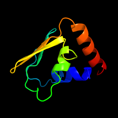
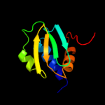
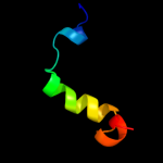
1 c1ibaA_
99.9
33
PDB header: phoshphotransferaseChain: A: PDB Molecule: glucose permease;PDBTitle: glucose permease (domain iib), nmr, 11 structures
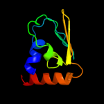
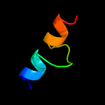
2 d3bp8c1
99.9
35
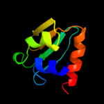
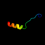
Fold: Homing endonuclease-likeSuperfamily: Glucose permease domain IIBFamily: Glucose permease domain IIB3 c3ipjB_
99.9
28
PDB header: transferaseChain: B: PDB Molecule: pts system, iiabc component;PDBTitle: the crystal structure of one domain of the pts system, iiabc component2 from clostridium difficile
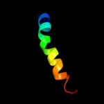
4 c2rddB_
72.7
17
PDB header: membrane protein/transport proteinChain: B: PDB Molecule: upf0092 membrane protein yajc;PDBTitle: x-ray crystal structure of acrb in complex with a novel2 transmembrane helix.
5 c3arct_
33.9
25
PDB header: electron transport, photosynthesisChain: T: PDB Molecule: photosystem ii reaction center protein t;PDBTitle: crystal structure of oxygen-evolving photosystem ii at 1.9 angstrom2 resolution
6 c3hd7A_
32.8
8
PDB header: exocytosisChain: A: PDB Molecule: vesicle-associated membrane protein 2;PDBTitle: helical extension of the neuronal snare complex into the membrane,2 spacegroup c 1 2 1
7 d3dtub2
26.2
12
Fold: Transmembrane helix hairpinSuperfamily: Cytochrome c oxidase subunit II-like, transmembrane regionFamily: Cytochrome c oxidase subunit II-like, transmembrane region8 c3mk7F_
19.0
26
PDB header: oxidoreductaseChain: F: PDB Molecule: cytochrome c oxidase, cbb3-type, subunit p;PDBTitle: the structure of cbb3 cytochrome oxidase
9 c2dlaB_
18.0
17
PDB header: replicationChain: B: PDB Molecule: 397aa long hypothetical protein;PDBTitle: primase large subunit amino terminal domain from pyrococcus horikoshii
10 c3a0bT_
16.4
25
PDB header: electron transportChain: T: PDB Molecule: photosystem ii reaction center protein t;PDBTitle: crystal structure of br-substituted photosystem ii complex
11 c1iijA_
12.8
19
PDB header: signaling proteinChain: A: PDB Molecule: erbb-2 receptor protein-tyrosine kinase;PDBTitle: solution structure of the neu/erbb-2 membrane spanning2 segment
12 d2cfua2
12.8
19
Fold: Metallo-hydrolase/oxidoreductaseSuperfamily: Metallo-hydrolase/oxidoreductaseFamily: Alkylsulfatase-like13 c2zjsE_
12.5
50
PDB header: protein transport/immune systemChain: E: PDB Molecule: preprotein translocase sece subunit;PDBTitle: crystal structure of secye translocon from thermus thermophilus with a2 fab fragment
14 c3tixB_
11.3
8
PDB header: gene regulation/protein bindingChain: B: PDB Molecule: chromo domain-containing protein 1;PDBTitle: crystal structure of the chp1-tas3 complex core
15 d1fftb2
10.8
8
Fold: Transmembrane helix hairpinSuperfamily: Cytochrome c oxidase subunit II-like, transmembrane regionFamily: Cytochrome c oxidase subunit II-like, transmembrane region16 c3bz1T_
10.4
25
PDB header: electron transportChain: T: PDB Molecule: photosystem ii reaction center protein t;PDBTitle: crystal structure of cyanobacterial photosystem ii (part 12 of 2). this file contains first monomer of psii dimer
17 c3prqT_
10.4
25
PDB header: photosynthesisChain: T: PDB Molecule: photosystem ii reaction center protein t;PDBTitle: crystal structure of cyanobacterial photosystem ii in complex with2 terbutryn (part 1 of 2). this file contains first monomer of psii3 dimer
18 c3bz2T_
10.4
25
PDB header: electron transportChain: T: PDB Molecule: photosystem ii reaction center protein t;PDBTitle: crystal structure of cyanobacterial photosystem ii (part 22 of 2). this file contains second monomer of psii dimer
19 c3prrT_
10.4
25
PDB header: photosynthesisChain: T: PDB Molecule: photosystem ii reaction center protein t;PDBTitle: crystal structure of cyanobacterial photosystem ii in complex with2 terbutryn (part 2 of 2). this file contains second monomer of psii3 dimer
20 c1s5lT_
10.3
25
PDB header: photosynthesisChain: T: PDB Molecule: photosystem ii psbt protein;PDBTitle: architecture of the photosynthetic oxygen evolving center
21 c1s5lt_
not modelled
10.3
25
PDB header: photosynthesisChain: T: PDB Molecule: photosystem ii psbt protein;PDBTitle: architecture of the photosynthetic oxygen evolving center
22 c3kziT_
not modelled
10.2
25
PDB header: electron transportChain: T: PDB Molecule: photosystem ii reaction center protein t;PDBTitle: crystal structure of monomeric form of cyanobacterial photosystem ii
23 c3arcT_
not modelled
10.2
25
PDB header: electron transport, photosynthesisChain: T: PDB Molecule: photosystem ii reaction center protein t;PDBTitle: crystal structure of oxygen-evolving photosystem ii at 1.9 angstrom2 resolution
24 d2axtt1
not modelled
10.2
25
Fold: Single transmembrane helixSuperfamily: Photosystem II reaction center protein T, PsbTFamily: PsbT-like25 c3a0bt_
not modelled
10.2
25
PDB header: electron transportChain: T: PDB Molecule: photosystem ii reaction center protein t;PDBTitle: crystal structure of br-substituted photosystem ii complex
26 c3a0ht_
not modelled
10.2
25
PDB header: electron transportChain: T: PDB Molecule: photosystem ii reaction center protein t;PDBTitle: crystal structure of i-substituted photosystem ii complex
27 c2axtT_
not modelled
10.2
25
PDB header: electron transportChain: T: PDB Molecule: photosystem ii reaction center t protein;PDBTitle: crystal structure of photosystem ii from thermosynechococcus elongatus
28 c2axtt_
not modelled
10.2
25
PDB header: electron transportChain: T: PDB Molecule: photosystem ii reaction center t protein;PDBTitle: crystal structure of photosystem ii from thermosynechococcus elongatus
29 c3a0hT_
not modelled
10.2
25
PDB header: electron transportChain: T: PDB Molecule: photosystem ii reaction center protein t;PDBTitle: crystal structure of i-substituted photosystem ii complex
30 c2cfuA_
not modelled
10.1
19
PDB header: hydrolaseChain: A: PDB Molecule: sdsa1;PDBTitle: crystal structure of sdsa1, an alkylsulfatase from2 pseudomonas aeruginosa, in complex with 1-decane-sulfonic-3 acid.
31 c3zqsB_
not modelled
8.0
17
PDB header: ligaseChain: B: PDB Molecule: e3 ubiquitin-protein ligase fancl;PDBTitle: human fancl central domain
32 c3d22A_
not modelled
6.9
14
PDB header: oxidoreductaseChain: A: PDB Molecule: thioredoxin h-type;PDBTitle: crystal structure of a poplar thioredoxin h mutant,2 pttrxh4c61s
33 d1fzda_
not modelled
6.7
22
Fold: Fibrinogen C-terminal domain-likeSuperfamily: Fibrinogen C-terminal domain-likeFamily: Fibrinogen C-terminal domain-like34 d1t57a_
not modelled
5.9
24
Fold: Pyruvate kinase C-terminal domain-likeSuperfamily: PK C-terminal domain-likeFamily: MTH1675-like35 c3qd1X_
not modelled
5.8
45
PDB header: sugar binding proteinChain: X: PDB Molecule: platelet binding protein gspb;PDBTitle: gspb plus alpha-2,3-sialyl (1-thioethyl)galactose
36 d3pvia_
not modelled
5.5
30
Fold: Restriction endonuclease-likeSuperfamily: Restriction endonuclease-likeFamily: Restriction endonuclease PvuII