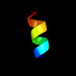
| Secondary structure and disorder prediction | |
| | |
1 | . | . | . | . | . | . | . | . | 10 | . | . | . | . | . | . | . | . | . | 20 | . | . | . | . | . | . | . | . | . | 30 | . | . | . | . | . | . | . | . | . | 40 | . | . | . | . | . | . | . |
| Sequence | |
M | K | K | F | R | W | V | V | L | V | V | V | V | L | A | C | L | L | L | W | A | Q | V | F | N | M | M | C | D | Q | D | V | Q | F | F | S | G | I | C | A | I | N | Q | F | I | P | W |
| Secondary structure | |
|  | | | | | | | | | | | | | | | | | | | | | | | | | | | | | | | | | | | | | | | | | | |
|
|
|
| SS confidence | |
|
|
|
|
|
|
|
|
|
|
|
|
|
|
|
|
|
|
|
|
|
|
|
|
|
|
|
|
|
|
|
|
|
|
|
|
|
|
|
|
|
|
|
|
|
|
|
| Disorder | |
? | ? | ? | ? |
|
|
|
|
|
|
|
|
|
|
|
|
|
|
|
|
|
|
|
|
|
|
|
|
|
|
|
|
|
|
|
|
|
|
|
|
|
|
|
|
| ? | ? |
| Disorder confidence | |
|
|
|
|
|
|
|
|
|
|
|
|
|
|
|
|
|
|
|
|
|
|
|
|
|
|
|
|
|
|
|
|
|
|
|
|
|
|
|
|
|
|
|
|
|
|
|
| |
| Confidence Key |
| High(9) | |
|
|
|
|
|
|
|
|
|
Low (0) |
| ? | Disordered |
| Alpha helix |
  | Beta strand |
Hover over an aligned region to see model and summary info
Please note, only up to the top 20 hits are modelled to reduce computer load
|
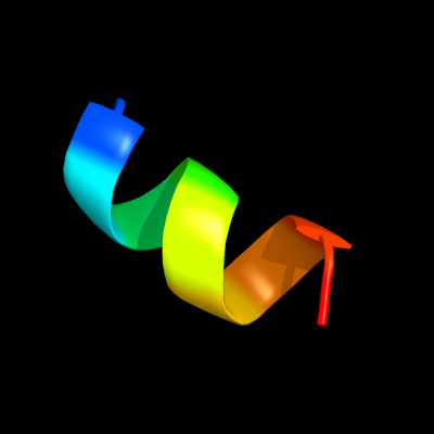
| 1 |
|
PDB 2bbn chain B
Region: 35 - 44
Aligned: 10
Modelled: 10
Confidence: 16.7%
Identity: 40%
PDB header:calcium-binding protein
Chain: B: PDB Molecule:myosin light chain kinase;
PDBTitle: solution structure of a calmodulin-target peptide complex2 by multidimensional nmr
Phyre2
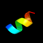
| 2 |
|
PDB 2bbm chain B
Region: 35 - 44
Aligned: 10
Modelled: 10
Confidence: 16.7%
Identity: 40%
PDB header:calcium-binding protein
Chain: B: PDB Molecule:myosin light chain kinase;
PDBTitle: solution structure of a calmodulin-target peptide complex2 by multidimensional nmr
Phyre2
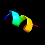
| 3 |
|
PDB 3bq9 chain A
Region: 33 - 42
Aligned: 10
Modelled: 10
Confidence: 10.0%
Identity: 50%
PDB header:structural genomics, unknown function
Chain: A: PDB Molecule:predicted rossmann fold nucleotide-binding domain-
PDBTitle: crystal structure of predicted nucleotide-binding protein from2 idiomarina baltica os145
Phyre2
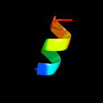
| 4 |
|
PDB 3gh1 chain A
Region: 33 - 42
Aligned: 10
Modelled: 10
Confidence: 9.7%
Identity: 50%
PDB header:structural genomics, unknown function
Chain: A: PDB Molecule:predicted nucleotide-binding protein;
PDBTitle: crystal structure of predicted nucleotide-binding protein from vibrio2 cholerae
Phyre2
|
| Detailed template information | |
Due to computational demand, binding site predictions are not run for batch jobs
If you want to predict binding sites, please manually submit your model of choice to 3DLigandSite
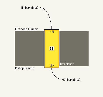
| Transmembrane helix prediction | |
Transmembrane helices have been predicted in your sequence to adopt the topology shown below
Phyre is for academic use only
| Please cite: Protein structure prediction on
the web: a case study using the Phyre server |
| Kelley LA and Sternberg MJE. Nature Protocols
4, 363 - 371 (2009) [pdf] [Import into BibTeX] |
| |
| If you use the binding site
predictions from 3DLigandSite, please also cite: |
| 3DLigandSite: predicting ligand-binding sites using similar structures. |
| Wass MN, Kelley LA and Sternberg
MJ Nucleic Acids Research 38, W469-73 (2010) [PubMed] |
| |
|
|
|
|