| 1 |
|
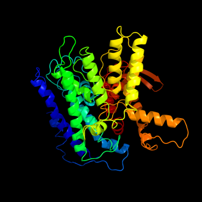
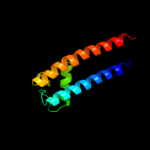
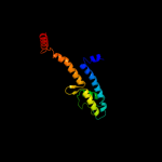
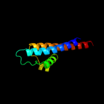
PDB 3pjz chain A
Region: 1 - 503
Aligned: 442
Modelled: 471
Confidence: 100.0%
Identity: 14%
PDB header:transport protein
Chain: A: PDB Molecule:potassium uptake protein trkh;
PDBTitle: crystal structure of the potassium transporter trkh from vibrio2 parahaemolyticus
Phyre2
| 2 |
|
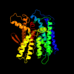
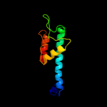
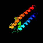
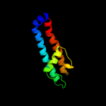
PDB 2qks chain A
Region: 413 - 513
Aligned: 98
Modelled: 101
Confidence: 92.4%
Identity: 16%
PDB header:metal transport
Chain: A: PDB Molecule:kir3.1-prokaryotic kir channel chimera;
PDBTitle: crystal structure of a kir3.1-prokaryotic kir channel chimera
Phyre2
| 3 |
|
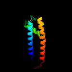
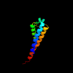
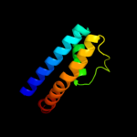
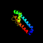
PDB 1xl4 chain A domain 2
Region: 413 - 511
Aligned: 96
Modelled: 99
Confidence: 91.3%
Identity: 15%
Fold: Voltage-gated potassium channels
Superfamily: Voltage-gated potassium channels
Family: Voltage-gated potassium channels
Phyre2
| 4 |
|
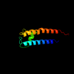
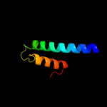
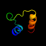
PDB 3jyc chain A
Region: 268 - 369
Aligned: 100
Modelled: 102
Confidence: 89.6%
Identity: 13%
PDB header:metal transport
Chain: A: PDB Molecule:inward-rectifier k+ channel kir2.2;
PDBTitle: crystal structure of the eukaryotic strong inward-rectifier2 k+ channel kir2.2 at 3.1 angstrom resolution
Phyre2
| 5 |
|
PDB 2a9h chain A domain 1
Region: 415 - 513
Aligned: 93
Modelled: 99
Confidence: 89.3%
Identity: 17%
Fold: Voltage-gated potassium channels
Superfamily: Voltage-gated potassium channels
Family: Voltage-gated potassium channels
Phyre2
| 6 |
|
PDB 1xl6 chain B
Region: 413 - 514
Aligned: 99
Modelled: 102
Confidence: 89.0%
Identity: 14%
PDB header:metal transport
Chain: B: PDB Molecule:inward rectifier potassium channel;
PDBTitle: intermediate gating structure 2 of the inwardly rectifying k+ channel2 kirbac3.1
Phyre2
| 7 |
|
PDB 1p7b chain B
Region: 413 - 512
Aligned: 98
Modelled: 100
Confidence: 89.0%
Identity: 12%
PDB header:metal transport
Chain: B: PDB Molecule:integral membrane channel and cytosolic domains;
PDBTitle: crystal structure of an inward rectifier potassium channel
Phyre2
| 8 |
|
PDB 3ifx chain B
Region: 416 - 512
Aligned: 92
Modelled: 97
Confidence: 87.4%
Identity: 15%
PDB header:membrane protein
Chain: B: PDB Molecule:voltage-gated potassium channel;
PDBTitle: crystal structure of the spin-labeled kcsa mutant v48r1
Phyre2
| 9 |
|
PDB 1f6g chain A
Region: 264 - 422
Aligned: 132
Modelled: 142
Confidence: 85.3%
Identity: 18%
Fold: Voltage-gated potassium channels
Superfamily: Voltage-gated potassium channels
Family: Voltage-gated potassium channels
Phyre2
| 10 |
|
PDB 1p7b chain A domain 2
Region: 413 - 509
Aligned: 95
Modelled: 97
Confidence: 78.1%
Identity: 13%
Fold: Voltage-gated potassium channels
Superfamily: Voltage-gated potassium channels
Family: Voltage-gated potassium channels
Phyre2
| 11 |
|
PDB 3e8g chain B
Region: 415 - 511
Aligned: 86
Modelled: 88
Confidence: 76.1%
Identity: 16%
PDB header:membrane protein
Chain: B: PDB Molecule:potassium channel protein;
PDBTitle: crystal structure of the the open nak channel-na+/ca2+ complex
Phyre2
| 12 |
|
PDB 2kb1 chain A
Region: 423 - 511
Aligned: 86
Modelled: 89
Confidence: 74.6%
Identity: 10%
PDB header:membrane protein
Chain: A: PDB Molecule:wsk3;
PDBTitle: nmr studies of a channel protein without membrane:2 structure and dynamics of water-solubilized kcsa
Phyre2
| 13 |
|
PDB 2h8p chain C domain 1
Region: 276 - 346
Aligned: 54
Modelled: 54
Confidence: 72.5%
Identity: 17%
Fold: Voltage-gated potassium channels
Superfamily: Voltage-gated potassium channels
Family: Voltage-gated potassium channels
Phyre2
| 14 |
|
PDB 1lnq chain C
Region: 333 - 373
Aligned: 39
Modelled: 41
Confidence: 69.4%
Identity: 28%
PDB header:metal transport
Chain: C: PDB Molecule:potassium channel related protein;
PDBTitle: crystal structure of mthk at 3.3 a
Phyre2
| 15 |
|
PDB 1r3j chain C
Region: 415 - 510
Aligned: 91
Modelled: 96
Confidence: 66.0%
Identity: 15%
Fold: Voltage-gated potassium channels
Superfamily: Voltage-gated potassium channels
Family: Voltage-gated potassium channels
Phyre2
| 16 |
|
PDB 2r9r chain H
Region: 407 - 506
Aligned: 97
Modelled: 100
Confidence: 59.8%
Identity: 7%
PDB header:membrane protein, transport protein
Chain: H: PDB Molecule:paddle chimera voltage gated potassium channel kv1.2-kv2.1;
PDBTitle: shaker family voltage dependent potassium channel (kv1.2-kv2.1 paddle2 chimera channel) in association with beta subunit
Phyre2
| 17 |
|
PDB 3beh chain A
Region: 407 - 506
Aligned: 97
Modelled: 100
Confidence: 58.3%
Identity: 12%
PDB header:membrane protein
Chain: A: PDB Molecule:mll3241 protein;
PDBTitle: structure of a bacterial cyclic nucleotide regulated ion channel
Phyre2
| 18 |
|
PDB 1lnq chain A domain 2
Region: 333 - 369
Aligned: 35
Modelled: 37
Confidence: 45.5%
Identity: 29%
Fold: Voltage-gated potassium channels
Superfamily: Voltage-gated potassium channels
Family: Voltage-gated potassium channels
Phyre2
| 19 |
|
PDB 1orq chain C
Region: 412 - 510
Aligned: 96
Modelled: 99
Confidence: 16.0%
Identity: 10%
Fold: Voltage-gated potassium channels
Superfamily: Voltage-gated potassium channels
Family: Voltage-gated potassium channels
Phyre2