Phyre Home Retrieve Phyre Job Id
Emaill.a.kelley@imperial.ac.uk
DescriptionP0AEV7
DateThu Jan 5 11:24:22 GMT 2012
Unique Job ID4f3311e04e73af5a

Summary 

Top model
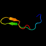
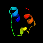
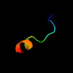
Image coloured by rainbow N → C terminus
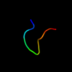
Model (left) based on template d1a6ca1
Top template information
Fold:Nucleoplasmin-like/VP (viral coat and capsid proteins)
Superfamily:Positive stranded ssRNA viruses
Family:Comoviridae-like VP
Confidence and coverage
Confidence: 70.4% Coverage: 20%
27 residues ( 20% of your sequence) have been modelled with 70.4% confidence by the single highest scoring template.
You may wish to submit your sequence to Phyrealarm. This will automatically scan your sequence every week for new potential templates as they appear in the Phyre2 library.
Please note: You must be registered and logged in to use Phyrealarm.
3D viewing
Interactive 3D view in Jmol

Sequence analysis 

Secondary structure and disorder prediction 

   1........10.........20.........30.........40.........50.........60
Sequence  MSEKVVFSQLSRKFIDENDATPAEAQQVVYYSLAIGHHLGVIDCLEAALTCPWDEYLAWI
Secondary structure 


SS confidence 


Disorder  ????


??????


Disorder confidence 


   .........70.........80.........90.........100.........110.........120
Sequence  ATLEAGSEARRKMEGVPKYGEIVIDINHVPMLANAFDKARAAQTSQQQEWSTMLLSMLHD
Secondary structure 


SS confidence 


Disorder 


??


???????


Disorder confidence 


   .........130......
Sequence  IHQENAIYLMVRRLRD
Secondary structure 


SS confidence 


Disorder 


?????
Disorder confidence 


Confidence Key
High(9)                    Low (0)
?Disordered
Alpha helix
Beta strand

Domain analysis 

Hover over an aligned region to see model and summary info

Please note, only up to the top 20 hits are modelled to reduce computer load

RankAligned region

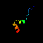
PDB 1a6c chain A domain 1

3D model

Region: 36 - 62
Aligned: 27
Modelled: 27
Confidence: 70.4%
Identity: 30%
Fold: Nucleoplasmin-like/VP (viral coat and capsid proteins)
Superfamily: Positive stranded ssRNA viruses
Family: Comoviridae-like VP

Phyre2

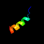
PDB 1wii chain A

3D model

Region: 49 - 66
Aligned: 18
Modelled: 18
Confidence: 26.8%
Identity: 44%
Fold: Rubredoxin-like
Superfamily: Zinc beta-ribbon
Family: Putative zinc binding domain

Phyre2

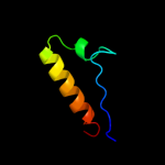
PDB 3e11 chain A domain 1

3D model

Region: 1 - 41
Aligned: 38
Modelled: 41
Confidence: 19.3%
Identity: 24%
Fold: Zincin-like
Superfamily: Metalloproteases ("zincins"), catalytic domain
Family: TTHA0227-like

Phyre2

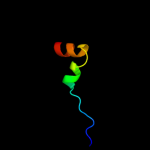
PDB 1a6c chain A

3D model

Region: 36 - 62
Aligned: 27
Modelled: 27
Confidence: 16.2%
Identity: 30%
PDB header:virus
Chain: A: PDB Molecule:tobacco ringspot virus capsid protein;
PDBTitle: structure of tobacco ringspot virus

Phyre2

PDB 2gk9 chain D

3D model

Region: 24 - 45
Aligned: 22
Modelled: 22
Confidence: 16.1%
Identity: 41%
PDB header:transferase
Chain: D: PDB Molecule:phosphatidylinositol-4-phosphate 5-kinase, type
PDBTitle: human phosphatidylinositol-4-phosphate 5-kinase, type ii,2 gamma

Phyre2

PDB 2y7u chain M

3D model

Region: 36 - 62
Aligned: 27
Modelled: 27
Confidence: 13.8%
Identity: 19%
PDB header:virus
Chain: M: PDB Molecule:coat protein;
PDBTitle: x-ray structure of the grapevine fanleaf virus

Phyre2

PDB 2q9l chain A

3D model

Region: 16 - 41
Aligned: 26
Modelled: 26
Confidence: 13.8%
Identity: 27%
PDB header:hydrolase
Chain: A: PDB Molecule:hypothetical protein;
PDBTitle: crystal structure of imazg from vibrio dat 722: ctag-imazg (p43212)

Phyre2

PDB 3dl8 chain D

3D model

Region: 28 - 43
Aligned: 16
Modelled: 16
Confidence: 12.6%
Identity: 50%
PDB header:protein transport
Chain: D: PDB Molecule:sece;
PDBTitle: structure of the complex of aquifex aeolicus secyeg and2 bacillus subtilis seca

Phyre2

PDB 1dxq chain A

3D model

Region: 31 - 40
Aligned: 10
Modelled: 10
Confidence: 10.6%
Identity: 60%
Fold: Flavodoxin-like
Superfamily: Flavoproteins
Family: Quinone reductase

Phyre2

PDB 3obc chain B

3D model

Region: 16 - 50
Aligned: 34
Modelled: 35
Confidence: 9.7%
Identity: 21%
PDB header:hydrolase
Chain: B: PDB Molecule:pyrophosphatase;
PDBTitle: crystal structure of a pyrophosphatase (af1178) from archaeoglobus2 fulgidus at 1.80 a resolution

Phyre2

PDB 2khs chain B

3D model

Region: 52 - 81
Aligned: 26
Modelled: 30
Confidence: 9.6%
Identity: 27%
PDB header:hydrolase
Chain: B: PDB Molecule:nuclease;
PDBTitle: solution structure of snase121:snase(111-143) complex

Phyre2

PDB 2oie chain A domain 1

3D model

Region: 16 - 41
Aligned: 26
Modelled: 26
Confidence: 8.6%
Identity: 27%
Fold: all-alpha NTP pyrophosphatases
Superfamily: all-alpha NTP pyrophosphatases
Family: MazG-like

Phyre2

PDB 2ejq chain A domain 1

3D model

Region: 1 - 39
Aligned: 37
Modelled: 39
Confidence: 8.4%
Identity: 19%
Fold: Zincin-like
Superfamily: Metalloproteases ("zincins"), catalytic domain
Family: TTHA0227-like

Phyre2

PDB 1ebf chain A domain 1

3D model

Region: 30 - 62
Aligned: 33
Modelled: 33
Confidence: 8.4%
Identity: 9%
Fold: NAD(P)-binding Rossmann-fold domains
Superfamily: NAD(P)-binding Rossmann-fold domains
Family: Glyceraldehyde-3-phosphate dehydrogenase-like, N-terminal domain

Phyre2

PDB 2gta chain D domain 1

3D model

Region: 16 - 41
Aligned: 26
Modelled: 26
Confidence: 8.3%
Identity: 12%
Fold: all-alpha NTP pyrophosphatases
Superfamily: all-alpha NTP pyrophosphatases
Family: MazG-like

Phyre2

PDB 2gta chain A domain 1

3D model

Region: 16 - 41
Aligned: 26
Modelled: 26
Confidence: 8.1%
Identity: 12%
Fold: all-alpha NTP pyrophosphatases
Superfamily: all-alpha NTP pyrophosphatases
Family: MazG-like

Phyre2

PDB 1vmg chain A

3D model

Region: 16 - 41
Aligned: 26
Modelled: 26
Confidence: 7.5%
Identity: 19%
Fold: all-alpha NTP pyrophosphatases
Superfamily: all-alpha NTP pyrophosphatases
Family: MazG-like

Phyre2

PDB 1cmc chain A

3D model

Region: 16 - 29
Aligned: 14
Modelled: 14
Confidence: 7.3%
Identity: 21%
Fold: Ribbon-helix-helix
Superfamily: Ribbon-helix-helix
Family: Met repressor, MetJ (MetR)

Phyre2

PDB 1qgp chain A

3D model

Region: 79 - 114
Aligned: 36
Modelled: 36
Confidence: 7.1%
Identity: 11%
Fold: DNA/RNA-binding 3-helical bundle
Superfamily: "Winged helix" DNA-binding domain
Family: Z-DNA binding domain

Phyre2

PDB 2q4p chain A

3D model

Region: 16 - 50
Aligned: 34
Modelled: 35
Confidence: 7.1%
Identity: 24%
PDB header:structural genomics, unknown function
Chain: A: PDB Molecule:protein rs21-c6;
PDBTitle: ensemble refinement of the crystal structure of protein from mus2 musculus mm.29898

Phyre2
1

d1a6ca1
2

d1wiia_
3

d3e11a1
4

c1a6cA_
5

c2gk9D_
6

c2y7uM_
7

c2q9lA_
8

c3dl8D_
9

d1dxqa_
10

c3obcB_
11

c2khsB_
12

d2oiea1
13

d2ejqa1
14

d1ebfa1
15

d2gtad1
16

d2gtaa1
17

d1vmga_
18

d1cmca_
19

d1qgpa_
20

c2q4pA_
21


22


23


24


25


26


27


28


29


30


Detailed template information 

#
Template Alignment Coverage3D Model Confidence
% i.d. Template Information
1d1a6ca1


70.4 30 Fold:Nucleoplasmin-like/VP (viral coat and capsid proteins)
Superfamily:Positive stranded ssRNA viruses
Family:Comoviridae-like VP
2d1wiia_


26.8 44 Fold:Rubredoxin-like
Superfamily:Zinc beta-ribbon
Family:Putative zinc binding domain
3d3e11a1


19.3 24 Fold:Zincin-like
Superfamily:Metalloproteases ("zincins"), catalytic domain
Family:TTHA0227-like
4c1a6cA_


16.2 30 PDB header:virus
Chain: A: PDB Molecule:tobacco ringspot virus capsid protein;
PDBTitle: structure of tobacco ringspot virus
5c2gk9D_


16.1 41 PDB header:transferase
Chain: D: PDB Molecule:phosphatidylinositol-4-phosphate 5-kinase, type
PDBTitle: human phosphatidylinositol-4-phosphate 5-kinase, type ii,2 gamma
6c2y7uM_


13.8 19 PDB header:virus
Chain: M: PDB Molecule:coat protein;
PDBTitle: x-ray structure of the grapevine fanleaf virus
7c2q9lA_


13.8 27 PDB header:hydrolase
Chain: A: PDB Molecule:hypothetical protein;
PDBTitle: crystal structure of imazg from vibrio dat 722: ctag-imazg (p43212)
8c3dl8D_


12.6 50 PDB header:protein transport
Chain: D: PDB Molecule:sece;
PDBTitle: structure of the complex of aquifex aeolicus secyeg and2 bacillus subtilis seca
9d1dxqa_


10.6 60 Fold:Flavodoxin-like
Superfamily:Flavoproteins
Family:Quinone reductase
10c3obcB_


9.7 21 PDB header:hydrolase
Chain: B: PDB Molecule:pyrophosphatase;
PDBTitle: crystal structure of a pyrophosphatase (af1178) from archaeoglobus2 fulgidus at 1.80 a resolution
11c2khsB_


9.6 27 PDB header:hydrolase
Chain: B: PDB Molecule:nuclease;
PDBTitle: solution structure of snase121:snase(111-143) complex
12d2oiea1


8.6 27 Fold:all-alpha NTP pyrophosphatases
Superfamily:all-alpha NTP pyrophosphatases
Family:MazG-like
13d2ejqa1


8.4 19 Fold:Zincin-like
Superfamily:Metalloproteases ("zincins"), catalytic domain
Family:TTHA0227-like
14d1ebfa1


8.4 9 Fold:NAD(P)-binding Rossmann-fold domains
Superfamily:NAD(P)-binding Rossmann-fold domains
Family:Glyceraldehyde-3-phosphate dehydrogenase-like, N-terminal domain
15d2gtad1


8.3 12 Fold:all-alpha NTP pyrophosphatases
Superfamily:all-alpha NTP pyrophosphatases
Family:MazG-like
16d2gtaa1


8.1 12 Fold:all-alpha NTP pyrophosphatases
Superfamily:all-alpha NTP pyrophosphatases
Family:MazG-like
17d1vmga_


7.5 19 Fold:all-alpha NTP pyrophosphatases
Superfamily:all-alpha NTP pyrophosphatases
Family:MazG-like
18d1cmca_


7.3 21 Fold:Ribbon-helix-helix
Superfamily:Ribbon-helix-helix
Family:Met repressor, MetJ (MetR)
19d1qgpa_


7.1 11 Fold:DNA/RNA-binding 3-helical bundle
Superfamily:"Winged helix" DNA-binding domain
Family:Z-DNA binding domain
20c2q4pA_


7.1 24 PDB header:structural genomics, unknown function
Chain: A: PDB Molecule:protein rs21-c6;
PDBTitle: ensemble refinement of the crystal structure of protein from mus2 musculus mm.29898
21d2a3qa1


not modelled 7.1 24 Fold:all-alpha NTP pyrophosphatases
Superfamily:all-alpha NTP pyrophosphatases
Family:MazG-like
22c2p7vA_


not modelled 6.9 24 PDB header:transcription
Chain: A: PDB Molecule:regulator of sigma d;
PDBTitle: crystal structure of the escherichia coli regulator of sigma 70, rsd,2 in complex with sigma 70 domain 4
23c3lj4i_


not modelled 6.9 20 PDB header:viral protein
Chain: I: PDB Molecule:portal protein;
PDBTitle: bacteriophage p22 portal protein bound to middle tail factor gp4. this2 file contain the first biological assembly
24d1xq5a_


not modelled 6.8 7 Fold:Globin-like
Superfamily:Globin-like
Family:Globins
25c2dzjA_


not modelled 6.0 33 PDB header:sugar binding protein
Chain: A: PDB Molecule:synaptic glycoprotein sc2;
PDBTitle: 2dzj/solution structure of the n-terminal ubiquitin-like2 domain in human synaptic glycoprotein sc2
26d2yzca2


not modelled 6.0 15 Fold:T-fold
Superfamily:Tetrahydrobiopterin biosynthesis enzymes-like
Family:Urate oxidase (uricase)
27c3efyB_


not modelled 6.0 15 PDB header:cell cycle
Chain: B: PDB Molecule:cif (cell cycle inhibiting factor);
PDBTitle: structure of the cyclomodulin cif from pathogenic2 escherichia coli
28d1pcfa_


not modelled 5.9 13 Fold:ssDNA-binding transcriptional regulator domain
Superfamily:ssDNA-binding transcriptional regulator domain
Family:Transcriptional coactivator PC4 C-terminal domain
29d1jl6a_


not modelled 5.6 13 Fold:Globin-like
Superfamily:Globin-like
Family:Globins
30c1otpA_


not modelled 5.2 24 PDB header:phosphorylase
Chain: A: PDB Molecule:thymidine phosphorylase;
PDBTitle: structural and theoretical studies suggest domain movement produces an2 active conformation of thymidine phosphorylase

Binding site prediction 

Due to computational demand, binding site predictions are not run for batch jobs

If you want to predict binding sites, please manually submit your model of choice to 3DLigandSite


Phyre is for academic use only

Please cite: Protein structure prediction on the web: a case study using the Phyre server
Kelley LA and Sternberg MJE. Nature Protocols 4, 363 - 371 (2009) [pdf] [Import into BibTeX]
 
If you use the binding site predictions from 3DLigandSite, please also cite:
3DLigandSite: predicting ligand-binding sites using similar structures.
Wass MN, Kelley LA and Sternberg MJ Nucleic Acids Research 38, W469-73 (2010) [PubMed]
 
© Structural Bioinformatics Group
Imperial College London
Lawrence Kelley, Benjamin Jefferys
Disclaimer
Terms and Conditions
Component software
Template detection: HHpred 1.51
Secondary structure prediction: Psi-pred 2.5
Disorder prediction: Disopred 2.4
Transmembrane prediction: Memsat_SVM
Multi-template modelling and ab initio: Poing 1.0