| 1 |
|
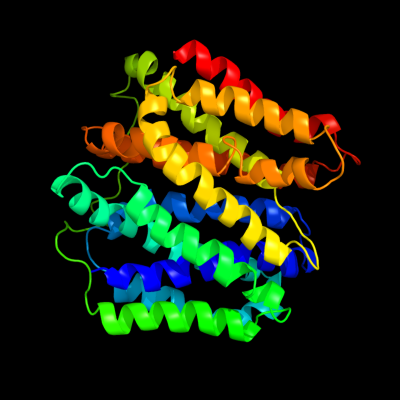
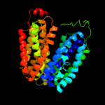
PDB 2gfp chain A
Region: 8 - 374
Aligned: 365
Modelled: 367
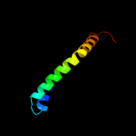
Confidence: 100.0%
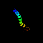
Identity: 21%
PDB header:membrane protein
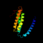
Chain: A: PDB Molecule:multidrug resistance protein d;
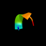
PDBTitle: structure of the multidrug transporter emrd from2 escherichia coli
Phyre2
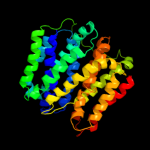
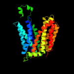
| 2 |
|
PDB 1pw4 chain A
Region: 5 - 395
Aligned: 390
Modelled: 391
Confidence: 100.0%
Identity: 13%
Fold: MFS general substrate transporter
Superfamily: MFS general substrate transporter
Family: Glycerol-3-phosphate transporter
Phyre2
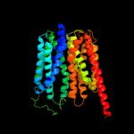
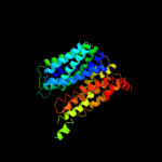
| 3 |
|
PDB 1pv7 chain A
Region: 4 - 397
Aligned: 394
Modelled: 394
Confidence: 100.0%
Identity: 10%
Fold: MFS general substrate transporter
Superfamily: MFS general substrate transporter
Family: LacY-like proton/sugar symporter
Phyre2
| 4 |
|
PDB 3o7p chain A
Region: 5 - 384
Aligned: 378
Modelled: 380
Confidence: 100.0%
Identity: 12%
PDB header:transport protein
Chain: A: PDB Molecule:l-fucose-proton symporter;
PDBTitle: crystal structure of the e.coli fucose:proton symporter, fucp (n162a)
Phyre2
| 5 |
|
PDB 2xut chain C
Region: 6 - 381
Aligned: 374
Modelled: 376
Confidence: 100.0%
Identity: 13%
PDB header:transport protein
Chain: C: PDB Molecule:proton/peptide symporter family protein;
PDBTitle: crystal structure of a proton dependent oligopeptide (pot)2 family transporter.
Phyre2
| 6 |
|
PDB 2g9p chain A
Region: 60 - 73
Aligned: 14
Modelled: 14
Confidence: 22.1%
Identity: 43%
PDB header:antimicrobial protein
Chain: A: PDB Molecule:antimicrobial peptide latarcin 2a;
PDBTitle: nmr structure of a novel antimicrobial peptide, latarcin 2a,2 from spider (lachesana tarabaevi) venom
Phyre2
| 7 |
|
PDB 2knc chain A
Region: 361 - 401
Aligned: 41
Modelled: 41
Confidence: 16.7%
Identity: 12%
PDB header:cell adhesion
Chain: A: PDB Molecule:integrin alpha-iib;
PDBTitle: platelet integrin alfaiib-beta3 transmembrane-cytoplasmic2 heterocomplex
Phyre2
| 8 |
|
PDB 3qnq chain D
Region: 348 - 400
Aligned: 53
Modelled: 53
Confidence: 16.5%
Identity: 6%
PDB header:membrane protein, transport protein
Chain: D: PDB Molecule:pts system, cellobiose-specific iic component;
PDBTitle: crystal structure of the transporter chbc, the iic component from the2 n,n'-diacetylchitobiose-specific phosphotransferase system
Phyre2
| 9 |
|
PDB 2xq2 chain A
Region: 263 - 403
Aligned: 136
Modelled: 136
Confidence: 7.5%
Identity: 7%
PDB header:transport protein
Chain: A: PDB Molecule:sodium/glucose cotransporter;
PDBTitle: structure of the k294a mutant of vsglt
Phyre2
| 10 |
|
PDB 3pro chain C domain 1
Region: 28 - 69
Aligned: 42
Modelled: 42
Confidence: 7.3%
Identity: 14%
Fold: Alpha-lytic protease prodomain-like
Superfamily: Alpha-lytic protease prodomain
Family: Alpha-lytic protease prodomain
Phyre2
| 11 |
|
PDB 1lzi chain A
Region: 399 - 403
Aligned: 5
Modelled: 5
Confidence: 5.9%
Identity: 80%
Fold: Nucleotide-diphospho-sugar transferases
Superfamily: Nucleotide-diphospho-sugar transferases
Family: alpha-1,3-galactosyltransferase-like
Phyre2