Phyre Home Retrieve Phyre Job Id
Emaill.a.kelley@imperial.ac.uk
DescriptionP76552
DateWed Jan 25 15:21:09 GMT 2012
Unique Job ID4d33e6cdac4bab53

Summary 

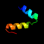
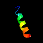
Top model
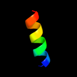
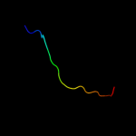
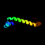
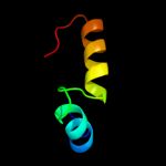
Image coloured by rainbow N → C terminus
Model (left) based on template d2hlya1
Top template information
Fold:Cysteine proteinases
Superfamily:Cysteine proteinases
Family:Atu2299-like
Confidence and coverage
Confidence: 35.4% Coverage: 4%
15 residues ( 4% of your sequence) have been modelled with 35.4% confidence by the single highest scoring template.
You may wish to submit your sequence to Phyrealarm. This will automatically scan your sequence every week for new potential templates as they appear in the Phyre2 library.
Please note: You must be registered and logged in to use Phyrealarm.
3D viewing
Interactive 3D view in Jmol

Sequence analysis 

Secondary structure and disorder prediction 

   1........10.........20.........30.........40.........50.........60
Sequence  MGINEIIMYIMMFFMLIAAVDRILSQFGGSARFLGKFGKSIEGSGGQFEEGFMAMGALGL
Secondary structure 


SS confidence 


Disorder  ??


????


?
??????


Disorder confidence 


   .........70.........80.........90.........100.........110.........120
Sequence  AMVGMTALAPVLAHVLGPVIIPVYEMLGANPSMFAGTLLACDMGGFFLAKELAGGDVAAW
Secondary structure 


SS confidence 


Disorder 


?
??


?


?????


Disorder confidence 


   .........130.........140.........150.........160.........170.........180
Sequence  LYSGLILGSMMGPTIVFSIPVALGIIEPSDRRYLALGVLAGIVTIPIGCIAGGLVAMYSG
Secondary structure 


SS confidence 


Disorder 


Disorder confidence 


   .........190.........200.........210.........220.........230.........240
Sequence  VQINGQPVEFTFALILMNMIPVIIVAILVALGLKFIPEKMINGFQIFAKFLVALITLGLA
Secondary structure 


SS confidence 


Disorder  ?
????????


Disorder confidence 


   .........250.........260.........270.........280.........290.........300
Sequence  AAVVKFLLGWELIPGLDPIFMAPGDKPGEVMRAIEVIGSISCVLLGAYPMVLLLTRWFEK
Secondary structure 


SS confidence 


Disorder 


?????


Disorder confidence 


   .........310.........320.........330.........340.........350.........360
Sequence  PLMSVGKVLNMNNIAAAGMVATLANNIPMFGMMKQMDTRGKVINCAFAVSAAFALGDHLG
Secondary structure 


SS confidence 


Disorder 


?


Disorder confidence 


   .........370.........380.........390.........400........
Sequence  FAAANMNAMIFPMIVGKLIGGVTAIGVAMMLVPKEDATATKTEAEAQS
Secondary structure 


SS confidence 


Disorder 


???
????
Disorder confidence 


Confidence Key
High(9)                    Low (0)
?Disordered
Alpha helix
Beta strand

Domain analysis 

Hover over an aligned region to see model and summary info

Please note, only up to the top 20 hits are modelled to reduce computer load

RankAligned region

PDB 2hly chain A domain 1

3D model

Region: 344 - 358
Aligned: 15
Modelled: 15
Confidence: 35.4%
Identity: 33%
Fold: Cysteine proteinases
Superfamily: Cysteine proteinases
Family: Atu2299-like

Phyre2

PDB 3qnq chain D

3D model

Region: 344 - 408
Aligned: 65
Modelled: 65
Confidence: 35.0%
Identity: 14%
PDB header:membrane protein, transport protein
Chain: D: PDB Molecule:pts system, cellobiose-specific iic component;
PDBTitle: crystal structure of the transporter chbc, the iic component from the2 n,n'-diacetylchitobiose-specific phosphotransferase system

Phyre2

PDB 3pjz chain A

3D model

Region: 255 - 407
Aligned: 150
Modelled: 153
Confidence: 34.3%
Identity: 14%
PDB header:transport protein
Chain: A: PDB Molecule:potassium uptake protein trkh;
PDBTitle: crystal structure of the potassium transporter trkh from vibrio2 parahaemolyticus

Phyre2

PDB 1j4n chain A

3D model

Region: 337 - 405
Aligned: 60
Modelled: 69
Confidence: 21.8%
Identity: 22%
Fold: Aquaporin-like
Superfamily: Aquaporin-like
Family: Aquaporin-like

Phyre2

PDB 3ajv chain A

3D model

Region: 312 - 343
Aligned: 32
Modelled: 32
Confidence: 17.3%
Identity: 16%
PDB header:hydrolase
Chain: A: PDB Molecule:putative uncharacterized protein;
PDBTitle: splicing endonuclease from aeropyrum pernix

Phyre2

PDB 2ntx chain B

3D model

Region: 334 - 354
Aligned: 21
Modelled: 21
Confidence: 16.3%
Identity: 29%
PDB header:signaling protein
Chain: B: PDB Molecule:emb
cab41934.1;

Phyre2

PDB 3ai9 chain X

3D model

Region: 352 - 363
Aligned: 12
Modelled: 12
Confidence: 16.0%
Identity: 42%
PDB header:transferase
Chain: X: PDB Molecule:upf0217 protein mj1640;
PDBTitle: crystal structure of duf358 protein reveals a putative spout-class2 rrna methyltransferase

Phyre2

PDB 2ka2 chain A

3D model

Region: 196 - 213
Aligned: 18
Modelled: 18
Confidence: 15.2%
Identity: 33%
PDB header:membrane protein
Chain: A: PDB Molecule:bcl2/adenovirus e1b 19 kda protein-interacting
PDBTitle: solution nmr structure of bnip3 transmembrane peptide dimer2 in detergent micelles with his173-ser172 intermonomer3 hydrogen bond restraints

Phyre2

PDB 2ka2 chain B

3D model

Region: 196 - 213
Aligned: 18
Modelled: 18
Confidence: 15.2%
Identity: 33%
PDB header:membrane protein
Chain: B: PDB Molecule:bcl2/adenovirus e1b 19 kda protein-interacting
PDBTitle: solution nmr structure of bnip3 transmembrane peptide dimer2 in detergent micelles with his173-ser172 intermonomer3 hydrogen bond restraints

Phyre2

PDB 2ka1 chain B

3D model

Region: 196 - 213
Aligned: 18
Modelled: 18
Confidence: 15.2%
Identity: 33%
PDB header:membrane protein
Chain: B: PDB Molecule:bcl2/adenovirus e1b 19 kda protein-interacting
PDBTitle: solution nmr structure of bnip3 transmembrane peptide dimer2 in detergent micelles

Phyre2

PDB 2ka1 chain A

3D model

Region: 196 - 213
Aligned: 18
Modelled: 18
Confidence: 15.2%
Identity: 33%
PDB header:membrane protein
Chain: A: PDB Molecule:bcl2/adenovirus e1b 19 kda protein-interacting
PDBTitle: solution nmr structure of bnip3 transmembrane peptide dimer2 in detergent micelles

Phyre2

PDB 2qwv chain A domain 1

3D model

Region: 352 - 363
Aligned: 12
Modelled: 12
Confidence: 15.0%
Identity: 25%
Fold: alpha/beta knot
Superfamily: alpha/beta knot
Family: AF1056-like

Phyre2

PDB 2qmm chain A domain 1

3D model

Region: 342 - 363
Aligned: 22
Modelled: 22
Confidence: 13.6%
Identity: 27%
Fold: alpha/beta knot
Superfamily: alpha/beta knot
Family: AF1056-like

Phyre2

PDB 1acc chain A

3D model

Region: 25 - 43
Aligned: 19
Modelled: 19
Confidence: 13.1%
Identity: 16%
Fold: Anthrax protective antigen
Superfamily: Anthrax protective antigen
Family: Anthrax protective antigen

Phyre2

PDB 1ig8 chain A domain 1

3D model

Region: 96 - 105
Aligned: 10
Modelled: 10
Confidence: 11.7%
Identity: 60%
Fold: Ribonuclease H-like motif
Superfamily: Actin-like ATPase domain
Family: Hexokinase

Phyre2

PDB 2j5d chain A

3D model

Region: 195 - 213
Aligned: 19
Modelled: 19
Confidence: 11.2%
Identity: 32%
PDB header:membrane protein
Chain: A: PDB Molecule:bcl2/adenovirus e1b 19 kda protein-interacting
PDBTitle: nmr structure of bnip3 transmembrane domain in lipid2 bicelles

Phyre2

PDB 1fx8 chain A

3D model

Region: 348 - 408
Aligned: 55
Modelled: 61
Confidence: 11.1%
Identity: 20%
Fold: Aquaporin-like
Superfamily: Aquaporin-like
Family: Aquaporin-like

Phyre2

PDB 1lda chain A

3D model

Region: 348 - 408
Aligned: 55
Modelled: 61
Confidence: 11.1%
Identity: 20%
PDB header:transport protein
Chain: A: PDB Molecule:glycerol uptake facilitator protein;
PDBTitle: crystal structure of the e. coli glycerol facilitator (glpf) without2 substrate glycerol

Phyre2

PDB 3zy6 chain A

3D model

Region: 101 - 136
Aligned: 28
Modelled: 36
Confidence: 11.1%
Identity: 29%
PDB header:transferase
Chain: A: PDB Molecule:putative gdp-fucose protein o-fucosyltransferase 1;
PDBTitle: crystal structure of pofut1 in complex with gdp-fucose2 (crystal-form-ii)

Phyre2

PDB 1cza chain N domain 1

3D model

Region: 96 - 105
Aligned: 10
Modelled: 10
Confidence: 10.8%
Identity: 50%
Fold: Ribonuclease H-like motif
Superfamily: Actin-like ATPase domain
Family: Hexokinase

Phyre2
1

d2hlya1
2

c3qnqD_
3

c3pjzA_
4

d1j4na_
5

c3ajvA_
6

c2ntxB_
7

c3ai9X_
8

c2ka2A_
9

c2ka2B_
10

c2ka1B_
11

c2ka1A_
12

d2qwva1
13

d2qmma1
14

d1acca_
15

d1ig8a1
16

c2j5dA_
17

d1fx8a_
18

c1ldaA_
19

c3zy6A_
20

d1czan1
21


22


23


24


25


26


27


28


29


30


31


32


33


34


35


36


37


38


39


40


41


42


43


44


45


46


47


48


49


50


51


52


53


54


Detailed template information 

#
Template Alignment Coverage3D Model Confidence
% i.d. Template Information
1d2hlya1


35.4 33 Fold:Cysteine proteinases
Superfamily:Cysteine proteinases
Family:Atu2299-like
2c3qnqD_


35.0 14 PDB header:membrane protein, transport protein
Chain: D: PDB Molecule:pts system, cellobiose-specific iic component;
PDBTitle: crystal structure of the transporter chbc, the iic component from the2 n,n'-diacetylchitobiose-specific phosphotransferase system
3c3pjzA_


34.3 14 PDB header:transport protein
Chain: A: PDB Molecule:potassium uptake protein trkh;
PDBTitle: crystal structure of the potassium transporter trkh from vibrio2 parahaemolyticus
4d1j4na_


21.8 22 Fold:Aquaporin-like
Superfamily:Aquaporin-like
Family:Aquaporin-like
5c3ajvA_


17.3 16 PDB header:hydrolase
Chain: A: PDB Molecule:putative uncharacterized protein;
PDBTitle: splicing endonuclease from aeropyrum pernix
6c2ntxB_


16.3 29 PDB header:signaling protein
Chain: B: PDB Molecule:emb
cab41934.1;
7c3ai9X_


16.0 42 PDB header:transferase
Chain: X: PDB Molecule:upf0217 protein mj1640;
PDBTitle: crystal structure of duf358 protein reveals a putative spout-class2 rrna methyltransferase
8c2ka2A_


15.2 33 PDB header:membrane protein
Chain: A: PDB Molecule:bcl2/adenovirus e1b 19 kda protein-interacting
PDBTitle: solution nmr structure of bnip3 transmembrane peptide dimer2 in detergent micelles with his173-ser172 intermonomer3 hydrogen bond restraints
9c2ka2B_


15.2 33 PDB header:membrane protein
Chain: B: PDB Molecule:bcl2/adenovirus e1b 19 kda protein-interacting
PDBTitle: solution nmr structure of bnip3 transmembrane peptide dimer2 in detergent micelles with his173-ser172 intermonomer3 hydrogen bond restraints
10c2ka1B_


15.2 33 PDB header:membrane protein
Chain: B: PDB Molecule:bcl2/adenovirus e1b 19 kda protein-interacting
PDBTitle: solution nmr structure of bnip3 transmembrane peptide dimer2 in detergent micelles
11c2ka1A_


15.2 33 PDB header:membrane protein
Chain: A: PDB Molecule:bcl2/adenovirus e1b 19 kda protein-interacting
PDBTitle: solution nmr structure of bnip3 transmembrane peptide dimer2 in detergent micelles
12d2qwva1


15.0 25 Fold:alpha/beta knot
Superfamily:alpha/beta knot
Family:AF1056-like
13d2qmma1


13.6 27 Fold:alpha/beta knot
Superfamily:alpha/beta knot
Family:AF1056-like
14d1acca_


13.1 16 Fold:Anthrax protective antigen
Superfamily:Anthrax protective antigen
Family:Anthrax protective antigen
15d1ig8a1


11.7 60 Fold:Ribonuclease H-like motif
Superfamily:Actin-like ATPase domain
Family:Hexokinase
16c2j5dA_


11.2 32 PDB header:membrane protein
Chain: A: PDB Molecule:bcl2/adenovirus e1b 19 kda protein-interacting
PDBTitle: nmr structure of bnip3 transmembrane domain in lipid2 bicelles
17d1fx8a_


11.1 20 Fold:Aquaporin-like
Superfamily:Aquaporin-like
Family:Aquaporin-like
18c1ldaA_


11.1 20 PDB header:transport protein
Chain: A: PDB Molecule:glycerol uptake facilitator protein;
PDBTitle: crystal structure of the e. coli glycerol facilitator (glpf) without2 substrate glycerol
19c3zy6A_


11.1 29 PDB header:transferase
Chain: A: PDB Molecule:putative gdp-fucose protein o-fucosyltransferase 1;
PDBTitle: crystal structure of pofut1 in complex with gdp-fucose2 (crystal-form-ii)
20d1czan1


10.8 50 Fold:Ribonuclease H-like motif
Superfamily:Actin-like ATPase domain
Family:Hexokinase
21c2b2hA_


not modelled 10.2 26 PDB header:transport protein
Chain: A: PDB Molecule:ammonium transporter;
PDBTitle: ammonium transporter amt-1 from a. fulgidus (as)
22c3op9A_


not modelled 10.0 14 PDB header:transcription regulator
Chain: A: PDB Molecule:pli0006 protein;
PDBTitle: crystal structure of transcriptional regulator from listeria innocua
23d1czan3


not modelled 9.9 60 Fold:Ribonuclease H-like motif
Superfamily:Actin-like ATPase domain
Family:Hexokinase
24d1bdga1


not modelled 9.3 60 Fold:Ribonuclease H-like motif
Superfamily:Actin-like ATPase domain
Family:Hexokinase
25d1bg3a3


not modelled 9.2 60 Fold:Ribonuclease H-like motif
Superfamily:Actin-like ATPase domain
Family:Hexokinase
26d1bg3a1


not modelled 9.1 50 Fold:Ribonuclease H-like motif
Superfamily:Actin-like ATPase domain
Family:Hexokinase
27c2k3uA_


not modelled 8.8 37 PDB header:immune system
Chain: A: PDB Molecule:chemotaxis inhibitory protein;
PDBTitle: structure of the tyrosine-sulfated c5a receptor n-terminus2 in complex with the immune evasion protein chips.
28c3llqB_


not modelled 8.8 19 PDB header:membrane protein
Chain: B: PDB Molecule:aquaporin z 2;
PDBTitle: aquaporin structure from plant pathogen agrobacterium tumerfaciens
29d1v4sa1


not modelled 8.4 50 Fold:Ribonuclease H-like motif
Superfamily:Actin-like ATPase domain
Family:Hexokinase
30c2lmfA_


not modelled 7.2 43 PDB header:antimicrobial protein
Chain: A: PDB Molecule:antibacterial protein ll-37;
PDBTitle: solution structure of human ll-23 bound to membrane-mimetic micelles
31c3hm8D_


not modelled 7.0 60 PDB header:transferase
Chain: D: PDB Molecule:hexokinase-3;
PDBTitle: crystal structure of the c-terminal hexokinase domain of human hk3
32c2w2eA_


not modelled 6.9 26 PDB header:membrane protein
Chain: A: PDB Molecule:aquaporin;
PDBTitle: 1.15 angstrom crystal structure of p.pastoris aquaporin,2 aqy1, in a closed conformation at ph 3.5
33d1rc2a_


not modelled 6.9 13 Fold:Aquaporin-like
Superfamily:Aquaporin-like
Family:Aquaporin-like
34c3p04B_


not modelled 6.6 14 PDB header:structural genomics, unknown function
Chain: B: PDB Molecule:uncharacterized bcr;
PDBTitle: crystal structure of the bcr protein from corynebacterium glutamicum.2 northeast structural genomics consortium target cgr8
35c2magA_


not modelled 6.4 35 PDB header:antibiotic
Chain: A: PDB Molecule:magainin 2;
PDBTitle: nmr structure of magainin 2 in dpc micelles, 10 structures
36c3c9jD_


not modelled 6.2 21 PDB header:membrane protein
Chain: D: PDB Molecule:proton channel protein m2, transmembrane segment;
PDBTitle: the crystal structure of transmembrane domain of m2 protein and2 amantadine complex
37c3c9jA_


not modelled 6.2 21 PDB header:membrane protein
Chain: A: PDB Molecule:proton channel protein m2, transmembrane segment;
PDBTitle: the crystal structure of transmembrane domain of m2 protein and2 amantadine complex
38c3c9jB_


not modelled 6.2 21 PDB header:membrane protein
Chain: B: PDB Molecule:proton channel protein m2, transmembrane segment;
PDBTitle: the crystal structure of transmembrane domain of m2 protein and2 amantadine complex
39c3c9jC_


not modelled 6.2 21 PDB header:membrane protein
Chain: C: PDB Molecule:proton channel protein m2, transmembrane segment;
PDBTitle: the crystal structure of transmembrane domain of m2 protein and2 amantadine complex
40c3ct5A_


not modelled 6.0 14 PDB header:hydrolase
Chain: A: PDB Molecule:morphogenesis protein 1;
PDBTitle: crystal and cryoem structural studies of a cell wall degrading enzyme2 in the bacteriophage phi29 tail
41d1u9ya2


not modelled 6.0 38 Fold:PRTase-like
Superfamily:PRTase-like
Family:Phosphoribosylpyrophosphate synthetase-like
42d1u7ga_


not modelled 5.9 16 Fold:Ammonium transporter
Superfamily:Ammonium transporter
Family:Ammonium transporter
43d1wi9a_


not modelled 5.7 63 Fold:DNA/RNA-binding 3-helical bundle
Superfamily:"Winged helix" DNA-binding domain
Family:PCI domain (PINT motif)
44c1ymgA_


not modelled 5.7 26 PDB header:membrane protein
Chain: A: PDB Molecule:lens fiber major intrinsic protein;
PDBTitle: the channel architecture of aquaporin o at 2.2 angstrom resolution
45d1ymga1


not modelled 5.7 26 Fold:Aquaporin-like
Superfamily:Aquaporin-like
Family:Aquaporin-like
46d1l8wd_


not modelled 5.6 35 Fold:Variable surface antigen VlsE
Superfamily:Variable surface antigen VlsE
Family:Variable surface antigen VlsE
47c3c1iA_


not modelled 5.5 20 PDB header:transport protein
Chain: A: PDB Molecule:ammonia channel;
PDBTitle: substrate binding, deprotonation and selectivity at the2 periplasmic entrance of the e. coli ammonia channel amtb
48c1iyjC_


not modelled 5.5 55 PDB header:gene regulation/antitumor protein
Chain: C: PDB Molecule:deleted in split hand/split foot protein 1;
PDBTitle: structure of a brca2-dss1 complex
49c1bdgA_


not modelled 5.5 60 PDB header:hexokinase
Chain: A: PDB Molecule:hexokinase;
PDBTitle: hexokinase from schistosoma mansoni complexed with glucose
50c1v4sA_


not modelled 5.5 50 PDB header:transferase
Chain: A: PDB Molecule:glucokinase isoform 2;
PDBTitle: crystal structure of human glucokinase
51c1ig8A_


not modelled 5.4 60 PDB header:transferase
Chain: A: PDB Molecule:hexokinase pii;
PDBTitle: crystal structure of yeast hexokinase pii with the correct2 amino acid sequence
52c2oarA_


not modelled 5.3 26 PDB header:membrane protein
Chain: A: PDB Molecule:large-conductance mechanosensitive channel;
PDBTitle: mechanosensitive channel of large conductance (mscl)
53c3hzqA_


not modelled 5.3 26 PDB header:membrane protein
Chain: A: PDB Molecule:large-conductance mechanosensitive channel;
PDBTitle: structure of a tetrameric mscl in an expanded intermediate2 state
54c3pvpA_


not modelled 5.1 10 PDB header:dna binding protein/dna
Chain: A: PDB Molecule:chromosomal replication initiator protein dnaa;
PDBTitle: structure of mycobacterium tuberculosis dnaa-dbd in complex with box22 dna

Binding site prediction 

Due to computational demand, binding site predictions are not run for batch jobs

If you want to predict binding sites, please manually submit your model of choice to 3DLigandSite


Phyre is for academic use only

Please cite: Protein structure prediction on the web: a case study using the Phyre server
Kelley LA and Sternberg MJE. Nature Protocols 4, 363 - 371 (2009) [pdf] [Import into BibTeX]
 
If you use the binding site predictions from 3DLigandSite, please also cite:
3DLigandSite: predicting ligand-binding sites using similar structures.
Wass MN, Kelley LA and Sternberg MJ Nucleic Acids Research 38, W469-73 (2010) [PubMed]
 
© Structural Bioinformatics Group
Imperial College London
Lawrence Kelley, Benjamin Jefferys
Disclaimer
Terms and Conditions
Component software
Template detection: HHpred 1.51
Secondary structure prediction: Psi-pred 2.5
Disorder prediction: Disopred 2.4
Transmembrane prediction: Memsat_SVM
Multi-template modelling and ab initio: Poing 1.0