Phyre Home Retrieve Phyre Job Id
Emaill.a.kelley@imperial.ac.uk
DescriptionP64530
DateThu Jan 5 12:09:14 GMT 2012
Unique Job ID4b5cab8747d5a7a4

Summary 

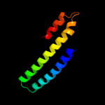
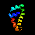
Top model
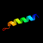
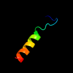
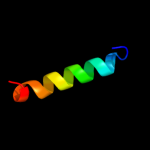
Image coloured by rainbow N → C terminus
Model (left) based on template c2hh7A_
Top template information
PDB header:unknown function
Chain: A: PDB Molecule:hypothetical protein csor;
PDBTitle: crystal structure of cu(i) bound csor from mycobacterium tuberculosis.
Confidence and coverage
Confidence: 99.9% Coverage: 91%
82 residues ( 91% of your sequence) have been modelled with 99.9% confidence by the single highest scoring template.
3D viewing
Interactive 3D view in Jmol

Sequence analysis 

Secondary structure and disorder prediction 

   1........10.........20.........30.........40.........50.........60
Sequence  MSHTIRDKQKLKARASKIQGQVVALKKMLDEPHECAAVLQQIAAIRGAVNGLMREVIKGH
Secondary structure 


SS confidence 


Disorder  ????


Disorder confidence 


   .........70.........80.........90
Sequence  LTEHIVHQGDELKREEDLDVVLKVLDSYIK
Secondary structure 


SS confidence 


Disorder 


???????


??
Disorder confidence 


Confidence Key
High(9)                    Low (0)
?Disordered
Alpha helix
Beta strand

Domain analysis 

Hover over an aligned region to see model and summary info

Please note, only up to the top 20 hits are modelled to reduce computer load

RankAligned region

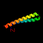
PDB 2hh7 chain A

3D model

Region: 5 - 86
Aligned: 82
Modelled: 82
Confidence: 99.9%
Identity: 16%
PDB header:unknown function
Chain: A: PDB Molecule:hypothetical protein csor;
PDBTitle: crystal structure of cu(i) bound csor from mycobacterium tuberculosis.

Phyre2

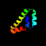
PDB 3aai chain B

3D model

Region: 1 - 74
Aligned: 74
Modelled: 71
Confidence: 99.9%
Identity: 19%
PDB header:transcription
Chain: B: PDB Molecule:copper homeostasis operon regulatory protein;
PDBTitle: x-ray crystal structure of csor from thermus thermophilus hb8

Phyre2

PDB 1paq chain A

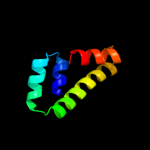
3D model

Region: 22 - 88
Aligned: 65
Modelled: 67
Confidence: 39.1%
Identity: 15%
PDB header:translation
Chain: A: PDB Molecule:translation initiation factor eif-2b epsilon
PDBTitle: crystal structure of the catalytic fragment of eukaryotic2 initiation factor 2b epsilon

Phyre2

PDB 1paq chain A

3D model

Region: 22 - 88
Aligned: 65
Modelled: 67
Confidence: 39.1%
Identity: 15%
Fold: alpha-alpha superhelix
Superfamily: ARM repeat
Family: MIF4G domain-like

Phyre2

PDB 3jui chain A

3D model

Region: 22 - 88
Aligned: 67
Modelled: 67
Confidence: 34.5%
Identity: 15%
PDB header:translation
Chain: A: PDB Molecule:translation initiation factor eif-2b subunit epsilon;
PDBTitle: crystal structure of the c-terminal domain of human translation2 initiation factor eif2b epsilon subunit

Phyre2

PDB 2p5t chain A

3D model

Region: 15 - 30
Aligned: 16
Modelled: 16
Confidence: 32.6%
Identity: 19%
PDB header:transcription regulator
Chain: A: PDB Molecule:putative transcriptional regulator peza;
PDBTitle: molecular and structural characterization of the pezat chromosomal2 toxin-antitoxin system of the human pathogen streptococcus pneumoniae

Phyre2

PDB 2nv2 chain U

3D model

Region: 10 - 43
Aligned: 34
Modelled: 34
Confidence: 25.1%
Identity: 21%
PDB header:lyase/transferase
Chain: U: PDB Molecule:pyridoxal biosynthesis lyase pdxs;
PDBTitle: structure of the plp synthase complex pdx1/2 (yaad/e) from bacillus2 subtilis

Phyre2

PDB 3zs9 chain D

3D model

Region: 4 - 21
Aligned: 18
Modelled: 18
Confidence: 16.5%
Identity: 50%
PDB header:hydrolase/transport protein
Chain: D: PDB Molecule:golgi to er traffic protein 2;
PDBTitle: s. cerevisiae get3-adp-alf4- complex with a cytosolic get2 fragment

Phyre2

PDB 2qyc chain A

3D model

Region: 4 - 25
Aligned: 22
Modelled: 22
Confidence: 12.2%
Identity: 5%
PDB header:unknown function
Chain: A: PDB Molecule:ferredoxin-like protein;
PDBTitle: crystal structure of a dimeric ferredoxin-like protein (bb1511) from2 bordetella bronchiseptica rb50 at 1.90 a resolution

Phyre2

PDB 1ivs chain A domain 1

3D model

Region: 5 - 36
Aligned: 32
Modelled: 32
Confidence: 11.7%
Identity: 9%
Fold: Long alpha-hairpin
Superfamily: tRNA-binding arm
Family: Valyl-tRNA synthetase (ValRS) C-terminal domain

Phyre2

PDB 3fem chain B

3D model

Region: 10 - 43
Aligned: 34
Modelled: 34
Confidence: 10.8%
Identity: 12%
PDB header:biosynthetic protein, transferase
Chain: B: PDB Molecule:pyridoxine biosynthesis protein snz1;
PDBTitle: structure of the synthase subunit pdx1.1 (snz1) of plp synthase from2 saccharomyces cerevisiae

Phyre2

PDB 3cve chain C

3D model

Region: 4 - 31
Aligned: 28
Modelled: 28
Confidence: 8.9%
Identity: 18%
PDB header:signaling protein
Chain: C: PDB Molecule:homer protein homolog 1;
PDBTitle: crystal structure of the carboxy terminus of homer1

Phyre2

PDB 2igs chain A domain 1

3D model

Region: 27 - 52
Aligned: 26
Modelled: 24
Confidence: 8.2%
Identity: 8%
Fold: Lysozyme-like
Superfamily: Lysozyme-like
Family: PA2222-like

Phyre2

PDB 1ev0 chain A

3D model

Region: 79 - 90
Aligned: 12
Modelled: 12
Confidence: 8.1%
Identity: 25%
Fold: Cell division protein MinE topological specificity domain
Superfamily: Cell division protein MinE topological specificity domain
Family: Cell division protein MinE topological specificity domain

Phyre2

PDB 1kkg chain A

3D model

Region: 2 - 24
Aligned: 23
Modelled: 23
Confidence: 8.0%
Identity: 4%
Fold: Alpha-lytic protease prodomain-like
Superfamily: Ribosome-binding factor A, RbfA
Family: Ribosome-binding factor A, RbfA

Phyre2

PDB 1obb chain A domain 2

3D model

Region: 38 - 90
Aligned: 51
Modelled: 53
Confidence: 8.0%
Identity: 16%
Fold: LDH C-terminal domain-like
Superfamily: LDH C-terminal domain-like
Family: AglA-like glucosidase

Phyre2

PDB 1jos chain A

3D model

Region: 2 - 23
Aligned: 22
Modelled: 22
Confidence: 7.2%
Identity: 9%
Fold: Alpha-lytic protease prodomain-like
Superfamily: Ribosome-binding factor A, RbfA
Family: Ribosome-binding factor A, RbfA

Phyre2

PDB 1tiz chain A

3D model

Region: 45 - 89
Aligned: 45
Modelled: 44
Confidence: 6.9%
Identity: 7%
Fold: EF Hand-like
Superfamily: EF-hand
Family: Calmodulin-like

Phyre2

PDB 3oq3 chain A

3D model

Region: 37 - 88
Aligned: 52
Modelled: 52
Confidence: 6.9%
Identity: 17%
PDB header:cytokine/viral protein
Chain: A: PDB Molecule:interferon alpha-5;
PDBTitle: structural basis of type-i interferon sequestration by a poxvirus2 decoy receptor

Phyre2

PDB 2ovc chain A

3D model

Region: 11 - 30
Aligned: 20
Modelled: 20
Confidence: 6.7%
Identity: 30%
PDB header:transport protein
Chain: A: PDB Molecule:potassium voltage-gated channel subfamily kqt member 4;
PDBTitle: crystal structure of a coiled-coil tetramerization domain from kv7.42 channels

Phyre2
1

c2hh7A_
2

c3aaiB_
3

c1paqA_
4

d1paqa_
5

c3juiA_
6

c2p5tA_
7

c2nv2U_
8

c3zs9D_
9

c2qycA_
10

d1ivsa1
11

c3femB_
12

c3cveC_
13

d2igsa1
14

d1ev0a_
15

d1kkga_
16

d1obba2
17

d1josa_
18

d1tiza_
19

c3oq3A_
20

c2ovcA_
21


22


23


24


25


26


27


Detailed template information 

#
Template Alignment Coverage3D Model Confidence
% i.d. Template Information
1c2hh7A_


99.9 16 PDB header:unknown function
Chain: A: PDB Molecule:hypothetical protein csor;
PDBTitle: crystal structure of cu(i) bound csor from mycobacterium tuberculosis.
2c3aaiB_


99.9 19 PDB header:transcription
Chain: B: PDB Molecule:copper homeostasis operon regulatory protein;
PDBTitle: x-ray crystal structure of csor from thermus thermophilus hb8
3c1paqA_


39.1 15 PDB header:translation
Chain: A: PDB Molecule:translation initiation factor eif-2b epsilon
PDBTitle: crystal structure of the catalytic fragment of eukaryotic2 initiation factor 2b epsilon
4d1paqa_


39.1 15 Fold:alpha-alpha superhelix
Superfamily:ARM repeat
Family:MIF4G domain-like
5c3juiA_


34.5 15 PDB header:translation
Chain: A: PDB Molecule:translation initiation factor eif-2b subunit epsilon;
PDBTitle: crystal structure of the c-terminal domain of human translation2 initiation factor eif2b epsilon subunit
6c2p5tA_


32.6 19 PDB header:transcription regulator
Chain: A: PDB Molecule:putative transcriptional regulator peza;
PDBTitle: molecular and structural characterization of the pezat chromosomal2 toxin-antitoxin system of the human pathogen streptococcus pneumoniae
7c2nv2U_


25.1 21 PDB header:lyase/transferase
Chain: U: PDB Molecule:pyridoxal biosynthesis lyase pdxs;
PDBTitle: structure of the plp synthase complex pdx1/2 (yaad/e) from bacillus2 subtilis
8c3zs9D_


16.5 50 PDB header:hydrolase/transport protein
Chain: D: PDB Molecule:golgi to er traffic protein 2;
PDBTitle: s. cerevisiae get3-adp-alf4- complex with a cytosolic get2 fragment
9c2qycA_


12.2 5 PDB header:unknown function
Chain: A: PDB Molecule:ferredoxin-like protein;
PDBTitle: crystal structure of a dimeric ferredoxin-like protein (bb1511) from2 bordetella bronchiseptica rb50 at 1.90 a resolution
10d1ivsa1


11.7 9 Fold:Long alpha-hairpin
Superfamily:tRNA-binding arm
Family:Valyl-tRNA synthetase (ValRS) C-terminal domain
11c3femB_


10.8 12 PDB header:biosynthetic protein, transferase
Chain: B: PDB Molecule:pyridoxine biosynthesis protein snz1;
PDBTitle: structure of the synthase subunit pdx1.1 (snz1) of plp synthase from2 saccharomyces cerevisiae
12c3cveC_


8.9 18 PDB header:signaling protein
Chain: C: PDB Molecule:homer protein homolog 1;
PDBTitle: crystal structure of the carboxy terminus of homer1
13d2igsa1


8.2 8 Fold:Lysozyme-like
Superfamily:Lysozyme-like
Family:PA2222-like
14d1ev0a_


8.1 25 Fold:Cell division protein MinE topological specificity domain
Superfamily:Cell division protein MinE topological specificity domain
Family:Cell division protein MinE topological specificity domain
15d1kkga_


8.0 4 Fold:Alpha-lytic protease prodomain-like
Superfamily:Ribosome-binding factor A, RbfA
Family:Ribosome-binding factor A, RbfA
16d1obba2


8.0 16 Fold:LDH C-terminal domain-like
Superfamily:LDH C-terminal domain-like
Family:AglA-like glucosidase
17d1josa_


7.2 9 Fold:Alpha-lytic protease prodomain-like
Superfamily:Ribosome-binding factor A, RbfA
Family:Ribosome-binding factor A, RbfA
18d1tiza_


6.9 7 Fold:EF Hand-like
Superfamily:EF-hand
Family:Calmodulin-like
19c3oq3A_


6.9 17 PDB header:cytokine/viral protein
Chain: A: PDB Molecule:interferon alpha-5;
PDBTitle: structural basis of type-i interferon sequestration by a poxvirus2 decoy receptor
20c2ovcA_


6.7 30 PDB header:transport protein
Chain: A: PDB Molecule:potassium voltage-gated channel subfamily kqt member 4;
PDBTitle: crystal structure of a coiled-coil tetramerization domain from kv7.42 channels
21d2oa5a1


not modelled 6.7 12 Fold:BLRF2-like
Superfamily:BLRF2-like
Family:BLRF2-like
22c2gtlO_


not modelled 6.2 21 PDB header:oxygen storage/transport
Chain: O: PDB Molecule:extracellular hemoglobin linker l3 subunit;
PDBTitle: lumbricus erythrocruorin at 3.5a resolution
23c1cffB_


not modelled 6.0 30 PDB header:calmodulin
Chain: B: PDB Molecule:protein (calcium pump);
PDBTitle: nmr solution structure of a complex of calmodulin with a2 binding peptide of the ca2+-pump
24c2kneB_


not modelled 6.0 31 PDB header:metal transport
Chain: B: PDB Molecule:atpase, ca++ transporting, plasma membrane 4;
PDBTitle: calmodulin wraps around its binding domain in the plasma2 membrane ca2+ pump anchored by a novel 18-1 motif
25d1up7a2


not modelled 5.8 16 Fold:LDH C-terminal domain-like
Superfamily:LDH C-terminal domain-like
Family:AglA-like glucosidase
26d1q4ra_


not modelled 5.5 5 Fold:Ferredoxin-like
Superfamily:Dimeric alpha+beta barrel
Family:Plant stress-induced protein
27c3nepX_


not modelled 5.1 21 PDB header:oxidoreductase
Chain: X: PDB Molecule:malate dehydrogenase;
PDBTitle: 1.55a resolution structure of malate dehydrogenase from salinibacter2 ruber

Binding site prediction 

Due to computational demand, binding site predictions are not run for batch jobs

If you want to predict binding sites, please manually submit your model of choice to 3DLigandSite


Phyre is for academic use only

Please cite: Protein structure prediction on the web: a case study using the Phyre server
Kelley LA and Sternberg MJE. Nature Protocols 4, 363 - 371 (2009) [pdf] [Import into BibTeX]
 
If you use the binding site predictions from 3DLigandSite, please also cite:
3DLigandSite: predicting ligand-binding sites using similar structures.
Wass MN, Kelley LA and Sternberg MJ Nucleic Acids Research 38, W469-73 (2010) [PubMed]
 
© Structural Bioinformatics Group
Imperial College London
Lawrence Kelley, Benjamin Jefferys
Disclaimer
Terms and Conditions
Component software
Template detection: HHpred 1.51
Secondary structure prediction: Psi-pred 2.5
Disorder prediction: Disopred 2.4
Transmembrane prediction: Memsat_SVM
Multi-template modelling and ab initio: Poing 1.0