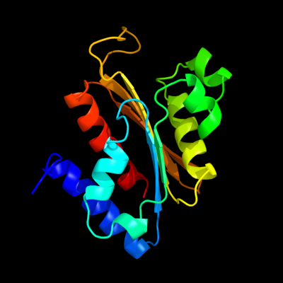
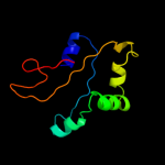
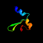
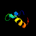
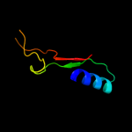
| 1 | d2a9sa1
|
|
 |
100.0 |
33 |
Fold:Anticodon-binding domain-like
Superfamily:CinA-like
Family:CinA-like |
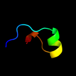
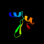
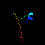
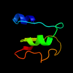
| 2 | d1pzna1
|
|
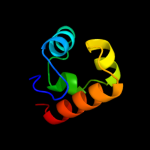 |
55.5 |
14 |
Fold:SAM domain-like
Superfamily:Rad51 N-terminal domain-like
Family:DNA repair protein Rad51, N-terminal domain |
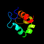
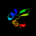
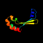
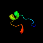
| 3 | d2i1qa1
|
|
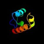 |
46.0 |
26 |
Fold:SAM domain-like
Superfamily:Rad51 N-terminal domain-like
Family:DNA repair protein Rad51, N-terminal domain |
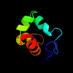
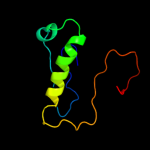
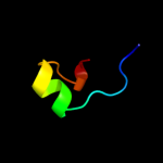
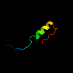
| 4 | c3b33A_
|
|
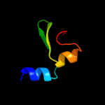 |
37.2 |
10 |
PDB header:transferase
Chain: A: PDB Molecule:sensor protein;
PDBTitle: crystal structure of the pas domain of nitrogen regulation protein2 nr(ii) from vibrio parahaemolyticus
|
| 5 | c3a9lB_
|
|
 |
36.1 |
14 |
PDB header:hydrolase
Chain: B: PDB Molecule:poly-gamma-glutamate hydrolase;
PDBTitle: structure of bacteriophage poly-gamma-glutamate hydrolase
|
| 6 | d1mzua_
|
|
 |
17.8 |
5 |
Fold:Profilin-like
Superfamily:PYP-like sensor domain (PAS domain)
Family:PYP-like |
| 7 | d1kfta_
|
|
 |
17.4 |
11 |
Fold:SAM domain-like
Superfamily:RuvA domain 2-like
Family:Excinuclease UvrC C-terminal domain |
| 8 | c1kftA_
|
|
 |
17.4 |
11 |
PDB header:dna binding protein
Chain: A: PDB Molecule:excinuclease abc subunit c;
PDBTitle: solution structure of the c-terminal domain of uvrc from e-2 coli
|
| 9 | d1v9ya_
|
|
 |
17.0 |
18 |
Fold:Profilin-like
Superfamily:PYP-like sensor domain (PAS domain)
Family:Heme-binding PAS domain |
| 10 | c1v9yA_
|
|
 |
17.0 |
18 |
PDB header:signaling protein
Chain: A: PDB Molecule:heme pas sensor protein;
PDBTitle: crystal structure of the heme pas sensor domain of ec dos (ferric2 form)
|
| 11 | c3mfxA_
|
|
 |
16.1 |
18 |
PDB header:transcription
Chain: A: PDB Molecule:sensory box/ggdef family protein;
PDBTitle: crystal structure of the sensory box domain of the sensory-2 box/ggdef protein so_1695 from shewanella oneidensis,3 northeast structural genomics consortium target sor288b
|
| 12 | d1saza1
|
|
 |
15.1 |
13 |
Fold:Ribonuclease H-like motif
Superfamily:Actin-like ATPase domain
Family:Acetokinase-like |
| 13 | c2o8kA_
|
|
 |
13.8 |
9 |
PDB header:transcription/dna
Chain: A: PDB Molecule:rna polymerase sigma factor rpon;
PDBTitle: nmr structure of the sigma-54 rpon domain bound to the-242 promoter element
|
| 14 | c3h3yF_
|
|
 |
12.4 |
22 |
PDB header:viral protein
Chain: F: PDB Molecule:baseplate structural protein gp6;
PDBTitle: fitting of the gp6 crystal structure into 3d cryo-em2 reconstruction of bacteriophage t4 star-shaped baseplate
|
| 15 | d1j3ma_
|
|
 |
12.2 |
15 |
Fold:TBP-like
Superfamily:TT1751-like
Family:TT1751-like |
| 16 | d1nwza_
|
|
 |
11.6 |
14 |
Fold:Profilin-like
Superfamily:PYP-like sensor domain (PAS domain)
Family:PYP-like |
| 17 | d2phna1
|
|
 |
11.0 |
29 |
Fold:CofE-like
Superfamily:CofE-like
Family:CofE-like |
| 18 | d1a4ia1
|
|
 |
10.6 |
31 |
Fold:NAD(P)-binding Rossmann-fold domains
Superfamily:NAD(P)-binding Rossmann-fold domains
Family:Aminoacid dehydrogenase-like, C-terminal domain |
| 19 | c3a0vA_
|
|
 |
10.4 |
10 |
PDB header:transferase
Chain: A: PDB Molecule:sensor protein;
PDBTitle: pas domain of histidine kinase thka (tm1359) (semet,2 f486m/f489m)
|
| 20 | c2w1tB_
|
|
 |
10.4 |
33 |
PDB header:transcription
Chain: B: PDB Molecule:stage v sporulation protein t;
PDBTitle: crystal structure of b. subtilis spovt
|
| 21 | d2bgwa1 |
|
not modelled |
9.8 |
10 |
Fold:SAM domain-like
Superfamily:RuvA domain 2-like
Family:Hef domain-like |
| 22 | c3mqoB_ |
|
not modelled |
9.7 |
11 |
PDB header:transcription regulator
Chain: B: PDB Molecule:transcriptional regulator, luxr family;
PDBTitle: the crystal structure of the pas domain in complex with isopropanol of2 a transcriptional regulator in the luxr family from burkholderia3 thailandensis to 1.7a
|
| 23 | c3ju7B_ |
|
not modelled |
9.6 |
7 |
PDB header:transferase
Chain: B: PDB Molecule:putative plp-dependent aminotransferase;
PDBTitle: crystal structure of putative plp-dependent aminotransferase2 (np_978343.1) from bacillus cereus atcc 10987 at 2.19 a resolution
|
| 24 | c3giuA_ |
|
not modelled |
8.9 |
31 |
PDB header:hydrolase
Chain: A: PDB Molecule:pyrrolidone-carboxylate peptidase;
PDBTitle: 1.25 angstrom crystal structure of pyrrolidone-carboxylate peptidase2 (pcp) from staphylococcus aureus
|
| 25 | d1xjca_ |
|
not modelled |
8.8 |
12 |
Fold:P-loop containing nucleoside triphosphate hydrolases
Superfamily:P-loop containing nucleoside triphosphate hydrolases
Family:Nitrogenase iron protein-like |
| 26 | c2v1bA_ |
|
not modelled |
8.8 |
15 |
PDB header:transferase
Chain: A: PDB Molecule:nph1-1;
PDBTitle: n- and c-terminal helices of oat lov2 (404-546) are2 involved in light-induced signal transduction (room3 temperature (293k) light structure of lov2 (404-546))
|
| 27 | c1zpdA_ |
|
not modelled |
8.4 |
17 |
PDB header:alcohol fermentation
Chain: A: PDB Molecule:pyruvate decarboxylase;
PDBTitle: pyruvate decarboxylase from zymomonas mobilis
|
| 28 | c1wu8B_ |
|
not modelled |
8.2 |
38 |
PDB header:structural genomics, unknown function
Chain: B: PDB Molecule:hypothetical protein ph0463;
PDBTitle: crystal structure of project ph0463 from pyrococcus horikoshii ot3
|
| 29 | c3p7nB_ |
|
not modelled |
8.1 |
13 |
PDB header:dna binding protein
Chain: B: PDB Molecule:sensor histidine kinase;
PDBTitle: crystal structure of light activated transcription factor el222 from2 erythrobacter litoralis
|
| 30 | c3fg8B_ |
|
not modelled |
7.9 |
10 |
PDB header:structural genomics, unknown function
Chain: B: PDB Molecule:uncharacterized protein rha05790;
PDBTitle: crystal structure of pas domain of rha05790
|
| 31 | d1otda_ |
|
not modelled |
7.8 |
14 |
Fold:Profilin-like
Superfamily:PYP-like sensor domain (PAS domain)
Family:PYP-like |
| 32 | c2zbvC_ |
|
not modelled |
7.5 |
31 |
PDB header:structural genomics, unknown function
Chain: C: PDB Molecule:uncharacterized conserved protein;
PDBTitle: crystal structure of uncharacterized conserved protein from thermotoga2 maritima
|
| 33 | d1rqpa2 |
|
not modelled |
7.5 |
31 |
Fold:Bacterial fluorinating enzyme, N-terminal domain
Superfamily:Bacterial fluorinating enzyme, N-terminal domain
Family:Bacterial fluorinating enzyme, N-terminal domain |
| 34 | d1iu8a_ |
|
not modelled |
7.2 |
50 |
Fold:Phosphorylase/hydrolase-like
Superfamily:Pyrrolidone carboxyl peptidase (pyroglutamate aminopeptidase)
Family:Pyrrolidone carboxyl peptidase (pyroglutamate aminopeptidase) |
| 35 | c3lacA_ |
|
not modelled |
7.0 |
44 |
PDB header:hydrolase
Chain: A: PDB Molecule:pyrrolidone-carboxylate peptidase;
PDBTitle: crystal structure of bacillus anthracis pyrrolidone-carboxylate2 peptidase, pcp
|
| 36 | c3gdwA_ |
|
not modelled |
6.7 |
15 |
PDB header:structural genomics, unknown function
Chain: A: PDB Molecule:sigma-54 interaction domain protein;
PDBTitle: crystal structure of sigma-54 interaction domain protein from2 enterococcus faecalis
|
| 37 | c2ebjB_ |
|
not modelled |
6.7 |
38 |
PDB header:hydrolase
Chain: B: PDB Molecule:pyrrolidone carboxyl peptidase;
PDBTitle: crystal structure of pyrrolidone carboxyl peptidase from thermus2 thermophilus
|
| 38 | c2v3wC_ |
|
not modelled |
6.1 |
16 |
PDB header:lyase
Chain: C: PDB Molecule:benzoylformate decarboxylase;
PDBTitle: crystal structure of the benzoylformate decarboxylase2 variant l461a from pseudomonas putida
|
| 39 | c3rtyA_ |
|
not modelled |
6.0 |
19 |
PDB header:circadian clock protein
Chain: A: PDB Molecule:period circadian protein;
PDBTitle: structure of an enclosed dimer formed by the drosophila period protein
|
| 40 | d1s2da_ |
|
not modelled |
5.8 |
11 |
Fold:Flavodoxin-like
Superfamily:N-(deoxy)ribosyltransferase-like
Family:N-deoxyribosyltransferase |
| 41 | d1iofa_ |
|
not modelled |
5.7 |
44 |
Fold:Phosphorylase/hydrolase-like
Superfamily:Pyrrolidone carboxyl peptidase (pyroglutamate aminopeptidase)
Family:Pyrrolidone carboxyl peptidase (pyroglutamate aminopeptidase) |
| 42 | c2cw5B_ |
|
not modelled |
5.6 |
18 |
PDB header:structural genomics, unknown function
Chain: B: PDB Molecule:bacterial fluorinating enzyme homolog;
PDBTitle: crystal structure of a conserved hypothetical protein from2 thermus thermophilus hb8
|
| 43 | c2q6oB_ |
|
not modelled |
5.5 |
38 |
PDB header:biosynthetic protein
Chain: B: PDB Molecule:hypothetical protein;
PDBTitle: sall-y70t with sam and cl
|
| 44 | c2e62A_ |
|
not modelled |
5.3 |
12 |
PDB header:rna binding protein
Chain: A: PDB Molecule:protein at5g25060;
PDBTitle: solution structure of the cwf21 domain in protein aak25922
|
| 45 | d1auga_ |
|
not modelled |
5.3 |
44 |
Fold:Phosphorylase/hydrolase-like
Superfamily:Pyrrolidone carboxyl peptidase (pyroglutamate aminopeptidase)
Family:Pyrrolidone carboxyl peptidase (pyroglutamate aminopeptidase) |
| 46 | c1rqrA_ |
|
not modelled |
5.2 |
31 |
PDB header:transferase
Chain: A: PDB Molecule:5'-fluoro-5'-deoxyadenosine synthase;
PDBTitle: crystal structure and mechanism of a bacterial fluorinating enzyme,2 product complex
|
| 47 | c2obxH_ |
|
not modelled |
5.2 |
11 |
PDB header:transferase
Chain: H: PDB Molecule:6,7-dimethyl-8-ribityllumazine synthase 1;
PDBTitle: lumazine synthase ribh2 from mesorhizobium loti (gene mll7281, swiss-2 prot entry q986n2) complexed with inhibitor 5-nitro-6-(d-3 ribitylamino)-2,4(1h,3h) pyrimidinedione
|
| 48 | c1hvwA_ |
|
not modelled |
5.0 |
100 |
PDB header:toxin
Chain: A: PDB Molecule:omega-atracotoxin-hv1a;
PDBTitle: hairpinless mutant of omega-atracotoxin-hv1a
|