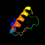
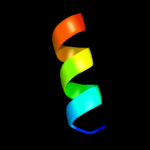
1 d1giqa2
15.6
24
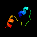
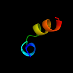
Fold: ADP-ribosylationSuperfamily: ADP-ribosylationFamily: ADP-ribosylating toxins2 d1fova_
11.6
43
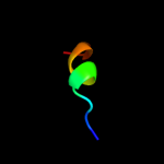
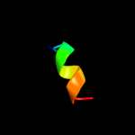
Fold: Thioredoxin foldSuperfamily: Thioredoxin-likeFamily: Thioltransferase3 d1jhba_
8.5
38
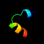
Fold: Thioredoxin foldSuperfamily: Thioredoxin-likeFamily: Thioltransferase4 c3l4nA_
8.0
31
PDB header: oxidoreductaseChain: A: PDB Molecule: monothiol glutaredoxin-6;PDBTitle: crystal structure of yeast monothiol glutaredoxin grx6
5 d1qs1a1
8.0
35
Fold: ADP-ribosylationSuperfamily: ADP-ribosylationFamily: ADP-ribosylating toxins6 c3fveA_
7.7
38
PDB header: isomeraseChain: A: PDB Molecule: diaminopimelate epimerase;PDBTitle: crystal structure of diaminopimelate epimerase mycobacterium2 tuberculosis dapf
7 d1t1va_
7.5
21
Fold: Thioredoxin foldSuperfamily: Thioredoxin-likeFamily: SH3BGR (SH3-binding, glutamic acid-rich protein-like)8 d1ejxc2
6.8
30
Fold: TIM beta/alpha-barrelSuperfamily: Metallo-dependent hydrolasesFamily: alpha-subunit of urease, catalytic domain9 d2hy5c1
6.6
22
Fold: DsrEFH-likeSuperfamily: DsrEFH-likeFamily: DsrH-like10 c3hymE_
6.6
70
PDB header: cell cycle, ligaseChain: E: PDB Molecule: anaphase-promoting complex subunit cdc26;PDBTitle: insights into anaphase promoting complex tpr subdomain2 assembly from a cdc26-apc6 structure
11 c3ogmB_
6.5
33
PDB header: protein bindingChain: B: PDB Molecule: coronatine-insensitive protein 1;PDBTitle: structure of coi1-ask1 in complex with coronatine and the jaz1 degron
12 d1ktea_
6.4
31
Fold: Thioredoxin foldSuperfamily: Thioredoxin-likeFamily: Thioltransferase13 c3oglD_
6.3
33
PDB header: protein bindingChain: D: PDB Molecule: coronatine-insensitive protein 1;PDBTitle: structure of coi1-ask1 in complex with ja-isoleucine and the jaz12 degron
14 c3hymK_
6.2
70
PDB header: cell cycle, ligaseChain: K: PDB Molecule: anaphase-promoting complex subunit cdc26;PDBTitle: insights into anaphase promoting complex tpr subdomain2 assembly from a cdc26-apc6 structure
15 c3ljbA_
6.2
12
PDB header: antiviral proteinChain: A: PDB Molecule: interferon-induced gtp-binding protein mx1;PDBTitle: structural basis of oligomerisation in the mxa stalk
16 d1qb5d_
6.1
60
Fold: OB-foldSuperfamily: Bacterial enterotoxinsFamily: Bacterial AB5 toxins, B-subunits17 c2wulB_
6.1
46
PDB header: oxidoreductaseChain: B: PDB Molecule: glutaredoxin related protein 5;PDBTitle: crystal structure of the human glutaredoxin 5 with bound2 glutathione in an fes cluster
18 d2gkea2
6.0
28
Fold: Diaminopimelate epimerase-likeSuperfamily: Diaminopimelate epimerase-likeFamily: Diaminopimelate epimerase19 c2hzfA_
5.9
31
PDB header: electron transport, oxidoreductaseChain: A: PDB Molecule: glutaredoxin-1;PDBTitle: crystal structures of a poxviral glutaredoxin in the oxidized and2 reduced states show redox-correlated structural changes
20 d1knya1
5.9
43
Fold: Four-helical up-and-down bundleSuperfamily: Nucleotidyltransferase substrate binding subunit/domainFamily: Kanamycin nucleotidyltransferase (KNTase), C-terminal domain21 c3hymA_
not modelled
5.9
70
PDB header: cell cycle, ligaseChain: A: PDB Molecule: anaphase-promoting complex subunit cdc26;PDBTitle: insights into anaphase promoting complex tpr subdomain2 assembly from a cdc26-apc6 structure
22 c3hymI_
not modelled
5.9
70
PDB header: cell cycle, ligaseChain: I: PDB Molecule: anaphase-promoting complex subunit cdc26;PDBTitle: insights into anaphase promoting complex tpr subdomain2 assembly from a cdc26-apc6 structure
23 c2ht9A_
not modelled
5.7
21
PDB header: oxidoreductaseChain: A: PDB Molecule: glutaredoxin-2;PDBTitle: the structure of dimeric human glutaredoxin 2
24 d2f6mb1
not modelled
5.4
29
Fold: Long alpha-hairpinSuperfamily: Endosomal sorting complex assembly domainFamily: VPS28 N-terminal domain25 d1p3qq_
not modelled
5.3
18
Fold: RuvA C-terminal domain-likeSuperfamily: UBA-likeFamily: CUE domain26 c3hymC_
not modelled
5.3
70
PDB header: cell cycle, ligaseChain: C: PDB Molecule: anaphase-promoting complex subunit cdc26;PDBTitle: insights into anaphase promoting complex tpr subdomain2 assembly from a cdc26-apc6 structure
27 d1r45a_
not modelled
5.3
29
Fold: ADP-ribosylationSuperfamily: ADP-ribosylationFamily: ADP-ribosylating toxins28 c2e7pC_
not modelled
5.2
31
PDB header: electron transportChain: C: PDB Molecule: glutaredoxin;PDBTitle: crystal structure of the holo form of glutaredoxin c1 from populus2 tremula x tremuloides
29 c2cazB_
not modelled
5.1
29
PDB header: protein transportChain: B: PDB Molecule: vacuolar protein sorting-associated proteinPDBTitle: escrt-i core
30 d2cazb1
not modelled
5.1
29
Fold: Long alpha-hairpinSuperfamily: Endosomal sorting complex assembly domainFamily: VPS28 N-terminal domain31 c3hymG_
not modelled
5.1
70
PDB header: cell cycle, ligaseChain: G: PDB Molecule: anaphase-promoting complex subunit cdc26;PDBTitle: insights into anaphase promoting complex tpr subdomain2 assembly from a cdc26-apc6 structure