Phyre Home Retrieve Phyre Job Id
Emaill.a.kelley@imperial.ac.uk
DescriptionP77453
DateThu Jan 5 12:29:24 GMT 2012
Unique Job ID4612cf2bffc669bd

Summary 

Top model
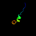
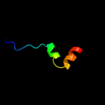
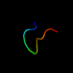
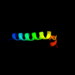
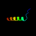
Image coloured by rainbow N → C terminus
Model (left) based on template d1a6ca1
Top template information
Fold:Nucleoplasmin-like/VP (viral coat and capsid proteins)
Superfamily:Positive stranded ssRNA viruses
Family:Comoviridae-like VP
Confidence and coverage
Confidence: 69.0% Coverage: 20%
27 residues ( 20% of your sequence) have been modelled with 69.0% confidence by the single highest scoring template.
You may wish to submit your sequence to Phyrealarm. This will automatically scan your sequence every week for new potential templates as they appear in the Phyre2 library.
Please note: You must be registered and logged in to use Phyrealarm.
3D viewing
Interactive 3D view in Jmol

Sequence analysis 

Secondary structure and disorder prediction 

   1........10.........20.........30.........40.........50.........60
Sequence  MTEECGEIVFWTLRKKFVASSDEMPEHSSQVMYYSLAIGHHVGVIDCLNVAFRCPLTEYE
Secondary structure 


SS confidence 


Disorder  ??????


??????


Disorder confidence 


   .........70.........80.........90.........100.........110.........120
Sequence  DWLALVEEEQARRKMLGVMTFGEIVIDASHTALLTRAFAPLADDATSVWQARSIQFIHLL
Secondary structure 


SS confidence 


Disorder 


?


????????


Disorder confidence 


   .........130.......
Sequence  DEIVQEPAIYLMARKIA
Secondary structure 


SS confidence 


Disorder 


???
Disorder confidence 


Confidence Key
High(9)                    Low (0)
?Disordered
Alpha helix
Beta strand

Domain analysis 

Hover over an aligned region to see model and summary info

Please note, only up to the top 20 hits are modelled to reduce computer load

RankAligned region

PDB 1a6c chain A domain 1

3D model

Region: 39 - 65
Aligned: 27
Modelled: 27
Confidence: 69.0%
Identity: 22%
Fold: Nucleoplasmin-like/VP (viral coat and capsid proteins)
Superfamily: Positive stranded ssRNA viruses
Family: Comoviridae-like VP

Phyre2

PDB 1wii chain A

3D model

Region: 52 - 69
Aligned: 18
Modelled: 18
Confidence: 20.5%
Identity: 28%
Fold: Rubredoxin-like
Superfamily: Zinc beta-ribbon
Family: Putative zinc binding domain

Phyre2

PDB 2p7v chain A

3D model

Region: 44 - 76
Aligned: 33
Modelled: 33
Confidence: 17.5%
Identity: 18%
PDB header:transcription
Chain: A: PDB Molecule:regulator of sigma d;
PDBTitle: crystal structure of the escherichia coli regulator of sigma 70, rsd,2 in complex with sigma 70 domain 4

Phyre2

PDB 2gk9 chain D

3D model

Region: 27 - 48
Aligned: 22
Modelled: 22
Confidence: 16.1%
Identity: 45%
PDB header:transferase
Chain: D: PDB Molecule:phosphatidylinositol-4-phosphate 5-kinase, type
PDBTitle: human phosphatidylinositol-4-phosphate 5-kinase, type ii,2 gamma

Phyre2

PDB 1a6c chain A

3D model

Region: 39 - 65
Aligned: 27
Modelled: 27
Confidence: 16.0%
Identity: 22%
PDB header:virus
Chain: A: PDB Molecule:tobacco ringspot virus capsid protein;
PDBTitle: structure of tobacco ringspot virus

Phyre2

PDB 2q9l chain A

3D model

Region: 19 - 44
Aligned: 26
Modelled: 26
Confidence: 16.0%
Identity: 27%
PDB header:hydrolase
Chain: A: PDB Molecule:hypothetical protein;
PDBTitle: crystal structure of imazg from vibrio dat 722: ctag-imazg (p43212)

Phyre2

PDB 3dl8 chain D

3D model

Region: 31 - 46
Aligned: 16
Modelled: 16
Confidence: 14.0%
Identity: 44%
PDB header:protein transport
Chain: D: PDB Molecule:sece;
PDBTitle: structure of the complex of aquifex aeolicus secyeg and2 bacillus subtilis seca

Phyre2

PDB 2y7u chain M

3D model

Region: 39 - 65
Aligned: 27
Modelled: 27
Confidence: 13.5%
Identity: 15%
PDB header:virus
Chain: M: PDB Molecule:coat protein;
PDBTitle: x-ray structure of the grapevine fanleaf virus

Phyre2

PDB 1dxq chain A

3D model

Region: 34 - 43
Aligned: 10
Modelled: 10
Confidence: 11.3%
Identity: 50%
Fold: Flavodoxin-like
Superfamily: Flavoproteins
Family: Quinone reductase

Phyre2

PDB 3aq0 chain G

3D model

Region: 15 - 81
Aligned: 63
Modelled: 67
Confidence: 11.3%
Identity: 11%
PDB header:transferase
Chain: G: PDB Molecule:geranyl diphosphate synthase;
PDBTitle: ligand-bound form of arabidopsis medium/long-chain length prenyl2 pyrophosphate synthase (surface polar residue mutant)

Phyre2

PDB 1cmc chain A

3D model

Region: 19 - 32
Aligned: 14
Modelled: 14
Confidence: 11.2%
Identity: 43%
Fold: Ribbon-helix-helix
Superfamily: Ribbon-helix-helix
Family: Met repressor, MetJ (MetR)

Phyre2

PDB 2id6 chain A domain 2

3D model

Region: 41 - 73
Aligned: 33
Modelled: 33
Confidence: 11.2%
Identity: 18%
Fold: Tetracyclin repressor-like, C-terminal domain
Superfamily: Tetracyclin repressor-like, C-terminal domain
Family: Tetracyclin repressor-like, C-terminal domain

Phyre2

PDB 3obc chain B

3D model

Region: 19 - 53
Aligned: 34
Modelled: 35
Confidence: 10.2%
Identity: 21%
PDB header:hydrolase
Chain: B: PDB Molecule:pyrophosphatase;
PDBTitle: crystal structure of a pyrophosphatase (af1178) from archaeoglobus2 fulgidus at 1.80 a resolution

Phyre2

PDB 2gta chain A domain 1

3D model

Region: 19 - 44
Aligned: 26
Modelled: 26
Confidence: 9.8%
Identity: 8%
Fold: all-alpha NTP pyrophosphatases
Superfamily: all-alpha NTP pyrophosphatases
Family: MazG-like

Phyre2

PDB 2oie chain A domain 1

3D model

Region: 19 - 44
Aligned: 26
Modelled: 26
Confidence: 9.6%
Identity: 23%
Fold: all-alpha NTP pyrophosphatases
Superfamily: all-alpha NTP pyrophosphatases
Family: MazG-like

Phyre2

PDB 3a9f chain A

3D model

Region: 5 - 34
Aligned: 30
Modelled: 30
Confidence: 9.5%
Identity: 10%
PDB header:electron transport
Chain: A: PDB Molecule:cytochrome c;
PDBTitle: crystal structure of the c-terminal domain of cytochrome cz2 from chlorobium tepidum

Phyre2

PDB 2gta chain D domain 1

3D model

Region: 19 - 44
Aligned: 26
Modelled: 26
Confidence: 9.4%
Identity: 8%
Fold: all-alpha NTP pyrophosphatases
Superfamily: all-alpha NTP pyrophosphatases
Family: MazG-like

Phyre2

PDB 2f3i chain A

3D model

Region: 118 - 137
Aligned: 20
Modelled: 20
Confidence: 9.2%
Identity: 25%
PDB header:transferase
Chain: A: PDB Molecule:dna-directed rna polymerases i, ii, and iii 17.1
PDBTitle: solution structure of a subunit of rna polymerase ii

Phyre2

PDB 1np3 chain A domain 1

3D model

Region: 81 - 132
Aligned: 52
Modelled: 52
Confidence: 8.7%
Identity: 21%
Fold: 6-phosphogluconate dehydrogenase C-terminal domain-like
Superfamily: 6-phosphogluconate dehydrogenase C-terminal domain-like
Family: Acetohydroxy acid isomeroreductase (ketol-acid reductoisomerase, KARI)

Phyre2

PDB 1vmg chain A

3D model

Region: 19 - 44
Aligned: 26
Modelled: 26
Confidence: 8.5%
Identity: 19%
Fold: all-alpha NTP pyrophosphatases
Superfamily: all-alpha NTP pyrophosphatases
Family: MazG-like

Phyre2
1

d1a6ca1
2

d1wiia_
3

c2p7vA_
4

c2gk9D_
5

c1a6cA_
6

c2q9lA_
7

c3dl8D_
8

c2y7uM_
9

d1dxqa_
10

c3aq0G_
11

d1cmca_
12

d2id6a2
13

c3obcB_
14

d2gtaa1
15

d2oiea1
16

c3a9fA_
17

d2gtad1
18

c2f3iA_
19

d1np3a1
20

d1vmga_
21


22


23


24


25


26


27


28


29


30


31


32


33


34


35


36


37


38


39


40


41


42


43


44


45


46


47


Detailed template information 

#
Template Alignment Coverage3D Model Confidence
% i.d. Template Information
1d1a6ca1


69.0 22 Fold:Nucleoplasmin-like/VP (viral coat and capsid proteins)
Superfamily:Positive stranded ssRNA viruses
Family:Comoviridae-like VP
2d1wiia_


20.5 28 Fold:Rubredoxin-like
Superfamily:Zinc beta-ribbon
Family:Putative zinc binding domain
3c2p7vA_


17.5 18 PDB header:transcription
Chain: A: PDB Molecule:regulator of sigma d;
PDBTitle: crystal structure of the escherichia coli regulator of sigma 70, rsd,2 in complex with sigma 70 domain 4
4c2gk9D_


16.1 45 PDB header:transferase
Chain: D: PDB Molecule:phosphatidylinositol-4-phosphate 5-kinase, type
PDBTitle: human phosphatidylinositol-4-phosphate 5-kinase, type ii,2 gamma
5c1a6cA_


16.0 22 PDB header:virus
Chain: A: PDB Molecule:tobacco ringspot virus capsid protein;
PDBTitle: structure of tobacco ringspot virus
6c2q9lA_


16.0 27 PDB header:hydrolase
Chain: A: PDB Molecule:hypothetical protein;
PDBTitle: crystal structure of imazg from vibrio dat 722: ctag-imazg (p43212)
7c3dl8D_


14.0 44 PDB header:protein transport
Chain: D: PDB Molecule:sece;
PDBTitle: structure of the complex of aquifex aeolicus secyeg and2 bacillus subtilis seca
8c2y7uM_


13.5 15 PDB header:virus
Chain: M: PDB Molecule:coat protein;
PDBTitle: x-ray structure of the grapevine fanleaf virus
9d1dxqa_


11.3 50 Fold:Flavodoxin-like
Superfamily:Flavoproteins
Family:Quinone reductase
10c3aq0G_


11.3 11 PDB header:transferase
Chain: G: PDB Molecule:geranyl diphosphate synthase;
PDBTitle: ligand-bound form of arabidopsis medium/long-chain length prenyl2 pyrophosphate synthase (surface polar residue mutant)
11d1cmca_


11.2 43 Fold:Ribbon-helix-helix
Superfamily:Ribbon-helix-helix
Family:Met repressor, MetJ (MetR)
12d2id6a2


11.2 18 Fold:Tetracyclin repressor-like, C-terminal domain
Superfamily:Tetracyclin repressor-like, C-terminal domain
Family:Tetracyclin repressor-like, C-terminal domain
13c3obcB_


10.2 21 PDB header:hydrolase
Chain: B: PDB Molecule:pyrophosphatase;
PDBTitle: crystal structure of a pyrophosphatase (af1178) from archaeoglobus2 fulgidus at 1.80 a resolution
14d2gtaa1


9.8 8 Fold:all-alpha NTP pyrophosphatases
Superfamily:all-alpha NTP pyrophosphatases
Family:MazG-like
15d2oiea1


9.6 23 Fold:all-alpha NTP pyrophosphatases
Superfamily:all-alpha NTP pyrophosphatases
Family:MazG-like
16c3a9fA_


9.5 10 PDB header:electron transport
Chain: A: PDB Molecule:cytochrome c;
PDBTitle: crystal structure of the c-terminal domain of cytochrome cz2 from chlorobium tepidum
17d2gtad1


9.4 8 Fold:all-alpha NTP pyrophosphatases
Superfamily:all-alpha NTP pyrophosphatases
Family:MazG-like
18c2f3iA_


9.2 25 PDB header:transferase
Chain: A: PDB Molecule:dna-directed rna polymerases i, ii, and iii 17.1
PDBTitle: solution structure of a subunit of rna polymerase ii
19d1np3a1


8.7 21 Fold:6-phosphogluconate dehydrogenase C-terminal domain-like
Superfamily:6-phosphogluconate dehydrogenase C-terminal domain-like
Family:Acetohydroxy acid isomeroreductase (ketol-acid reductoisomerase, KARI)
20d1vmga_


8.5 19 Fold:all-alpha NTP pyrophosphatases
Superfamily:all-alpha NTP pyrophosphatases
Family:MazG-like
21d1ebfa1


not modelled 8.2 12 Fold:NAD(P)-binding Rossmann-fold domains
Superfamily:NAD(P)-binding Rossmann-fold domains
Family:Glyceraldehyde-3-phosphate dehydrogenase-like, N-terminal domain
22c2khsB_


not modelled 7.9 27 PDB header:hydrolase
Chain: B: PDB Molecule:nuclease;
PDBTitle: solution structure of snase121:snase(111-143) complex
23c2l5fA_


not modelled 7.6 10 PDB header:protein binding
Chain: A: PDB Molecule:pre-mrna-processing factor 40 homolog a;
PDBTitle: solution structure of the tandem ww domains from hypa/fbp11
24c2q4pA_


not modelled 7.2 18 PDB header:structural genomics, unknown function
Chain: A: PDB Molecule:protein rs21-c6;
PDBTitle: ensemble refinement of the crystal structure of protein from mus2 musculus mm.29898
25d2a3qa1


not modelled 7.2 18 Fold:all-alpha NTP pyrophosphatases
Superfamily:all-alpha NTP pyrophosphatases
Family:MazG-like
26c3lj4i_


not modelled 7.0 10 PDB header:viral protein
Chain: I: PDB Molecule:portal protein;
PDBTitle: bacteriophage p22 portal protein bound to middle tail factor gp4. this2 file contain the first biological assembly
27c3iz5W_


not modelled 6.1 39 PDB header:ribosome
Chain: W: PDB Molecule:60s ribosomal protein l22 (l22e);
PDBTitle: localization of the large subunit ribosomal proteins into a 5.5 a2 cryo-em map of triticum aestivum translating 80s ribosome
28c3p7jA_


not modelled 6.0 16 PDB header:transcription
Chain: A: PDB Molecule:heterochromatin protein 1;
PDBTitle: drosophila hp1a chromo shadow domain
29d1pn2a1


not modelled 6.0 33 Fold:Thioesterase/thiol ester dehydrase-isomerase
Superfamily:Thioesterase/thiol ester dehydrase-isomerase
Family:MaoC-like
30d1d8ca_


not modelled 6.0 19 Fold:TIM beta/alpha-barrel
Superfamily:Malate synthase G
Family:Malate synthase G
31d1pcfa_


not modelled 5.9 13 Fold:ssDNA-binding transcriptional regulator domain
Superfamily:ssDNA-binding transcriptional regulator domain
Family:Transcriptional coactivator PC4 C-terminal domain
32d2rm0w1


not modelled 5.8 20 Fold:WW domain-like
Superfamily:WW domain
Family:WW domain
33d1unda_


not modelled 5.8 6 Fold:VHP, Villin headpiece domain
Superfamily:VHP, Villin headpiece domain
Family:VHP, Villin headpiece domain
34d2hanb1


not modelled 5.8 25 Fold:Glucocorticoid receptor-like (DNA-binding domain)
Superfamily:Glucocorticoid receptor-like (DNA-binding domain)
Family:Nuclear receptor
35c3oyrA_


not modelled 5.7 15 PDB header:transferase
Chain: A: PDB Molecule:trans-isoprenyl diphosphate synthase;
PDBTitle: crystal structure of polyprenyl synthase from caulobacter crescentus2 cb15 complexed with calcium and isoprenyl diphosphate
36d1gpca_


not modelled 5.6 24 Fold:OB-fold
Superfamily:Nucleic acid-binding proteins
Family:Phage ssDNA-binding proteins
37c2dzjA_


not modelled 5.5 33 PDB header:sugar binding protein
Chain: A: PDB Molecule:synaptic glycoprotein sc2;
PDBTitle: 2dzj/solution structure of the n-terminal ubiquitin-like2 domain in human synaptic glycoprotein sc2
38d1qzpa_


not modelled 5.4 12 Fold:VHP, Villin headpiece domain
Superfamily:VHP, Villin headpiece domain
Family:VHP, Villin headpiece domain
39c3efyB_


not modelled 5.4 19 PDB header:cell cycle
Chain: B: PDB Molecule:cif (cell cycle inhibiting factor);
PDBTitle: structure of the cyclomodulin cif from pathogenic2 escherichia coli
40c3gp2B_


not modelled 5.4 71 PDB header:metal binding protein/transferase
Chain: B: PDB Molecule:calcium/calmodulin-dependent protein kinase type
PDBTitle: calmodulin bound to peptide from calmodulin kinase ii2 (camkii)
41c1cm4B_


not modelled 5.4 71 PDB header:complex (calcium-binding/transferase)
Chain: B: PDB Molecule:calmodulin-dependent protein kinase ii-alpha;
PDBTitle: motions of calmodulin-four-conformer refinement
42c1cdmB_


not modelled 5.4 71 PDB header:calcium-binding protein
Chain: B: PDB Molecule:calmodulin;
PDBTitle: modulation of calmodulin plasticity in molecular2 recognition on the basis of x-ray structures
43c1cm1B_


not modelled 5.4 71 PDB header:complex (calcium-binding/transferase)
Chain: B: PDB Molecule:calmodulin-dependent protein kinase ii-alpha;
PDBTitle: motions of calmodulin-single-conformer refinement
44d2fmma1


not modelled 5.2 23 Fold:SH3-like barrel
Superfamily:Chromo domain-like
Family:Chromo domain
45d1thta_


not modelled 5.1 55 Fold:alpha/beta-Hydrolases
Superfamily:alpha/beta-Hydrolases
Family:Thioesterases
46d2o3bb1


not modelled 5.1 40 Fold:Nuclease A inhibitor (NuiA)
Superfamily:Nuclease A inhibitor (NuiA)
Family:Nuclease A inhibitor (NuiA)
47c3cbbA_


not modelled 5.0 31 PDB header:transcription/dna
Chain: A: PDB Molecule:hepatocyte nuclear factor 4-alpha, dna binding
PDBTitle: crystal structure of hepatocyte nuclear factor 4alpha in2 complex with dna: diabetes gene product

Binding site prediction 

Due to computational demand, binding site predictions are not run for batch jobs

If you want to predict binding sites, please manually submit your model of choice to 3DLigandSite


Phyre is for academic use only

Please cite: Protein structure prediction on the web: a case study using the Phyre server
Kelley LA and Sternberg MJE. Nature Protocols 4, 363 - 371 (2009) [pdf] [Import into BibTeX]
 
If you use the binding site predictions from 3DLigandSite, please also cite:
3DLigandSite: predicting ligand-binding sites using similar structures.
Wass MN, Kelley LA and Sternberg MJ Nucleic Acids Research 38, W469-73 (2010) [PubMed]
 
© Structural Bioinformatics Group
Imperial College London
Lawrence Kelley, Benjamin Jefferys
Disclaimer
Terms and Conditions
Component software
Template detection: HHpred 1.51
Secondary structure prediction: Psi-pred 2.5
Disorder prediction: Disopred 2.4
Transmembrane prediction: Memsat_SVM
Multi-template modelling and ab initio: Poing 1.0