1 d1kqfc_
99.9
16
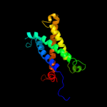
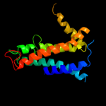
Fold: Heme-binding four-helical bundleSuperfamily: Transmembrane di-heme cytochromesFamily: Formate dehydrogenase N, cytochrome (gamma) subunit2 c2qjkM_
97.1
13
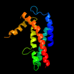
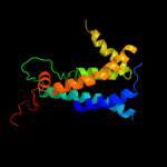
PDB header: electron transportChain: M: PDB Molecule: cytochrome b;PDBTitle: crystal structure analysis of mutant rhodobacter2 sphaeroides bc1 with stigmatellin and antimycin
3 d3cx5c2
97.0
12
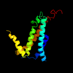
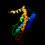
Fold: Heme-binding four-helical bundleSuperfamily: Transmembrane di-heme cytochromesFamily: Cytochrome b of cytochrome bc1 complex (Ubiquinol-cytochrome c reductase)4 c3cx5N_
97.0
14
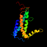
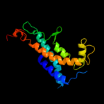
PDB header: oxidoreductaseChain: N: PDB Molecule: cytochrome b;PDBTitle: structure of complex iii with bound cytochrome c in reduced2 state and definition of a minimal core interface for3 electron transfer.
5 d1q90b_
96.9
18
Fold: Heme-binding four-helical bundleSuperfamily: Transmembrane di-heme cytochromesFamily: Cytochrome b of cytochrome bc1 complex (Ubiquinol-cytochrome c reductase)6 d1ppjc2
96.5
16
Fold: Heme-binding four-helical bundleSuperfamily: Transmembrane di-heme cytochromesFamily: Cytochrome b of cytochrome bc1 complex (Ubiquinol-cytochrome c reductase)7 c3cwbC_
96.5
16
PDB header: oxidoreductaseChain: C: PDB Molecule: cytochrome b;PDBTitle: chicken cytochrome bc1 complex inhibited by an iodinated analogue of2 the polyketide crocacin-d
8 d1bccc3
96.5
17
Fold: Heme-binding four-helical bundleSuperfamily: Transmembrane di-heme cytochromesFamily: Cytochrome b of cytochrome bc1 complex (Ubiquinol-cytochrome c reductase)9 d2e74a1
96.4
18
Fold: Heme-binding four-helical bundleSuperfamily: Transmembrane di-heme cytochromesFamily: Cytochrome b of cytochrome bc1 complex (Ubiquinol-cytochrome c reductase)10 d1y5ic1
67.7
14
Fold: Heme-binding four-helical bundleSuperfamily: Respiratory nitrate reductase 1 gamma chainFamily: Respiratory nitrate reductase 1 gamma chain11 d2ieca1
20.4
30
Fold: MK0786-likeSuperfamily: MK0786-likeFamily: MK0786-like12 c2ogfD_
17.5
40
PDB header: structural genomics, unknown functionChain: D: PDB Molecule: hypothetical protein mj0408;PDBTitle: crystal structure of protein mj0408 from methanococcus jannaschii,2 pfam duf372
13 d2i52a1
16.9
20
Fold: MK0786-likeSuperfamily: MK0786-likeFamily: MK0786-like14 d1t33a1
10.4
22
Fold: DNA/RNA-binding 3-helical bundleSuperfamily: Homeodomain-likeFamily: Tetracyclin repressor-like, N-terminal domain15 c3g56A_
7.6
39
PDB header: dna binding proteinChain: A: PDB Molecule: regulator of macrolide 2'-phosphotransferase i;PDBTitle: structure of the macrolide biosensor protein, mphr(a)
16 d1m56d_
7.4
21
Fold: Single transmembrane helixSuperfamily: Bacterial aa3 type cytochrome c oxidase subunit IVFamily: Bacterial aa3 type cytochrome c oxidase subunit IV17 c2yvhA_
7.2
22
PDB header: transcription/dnaChain: A: PDB Molecule: transcriptional regulator;PDBTitle: crystal structure of the operator-binding form of the multi-drug2 binding transcriptional repressor cgmr
18 c2hkuB_
6.9
7
PDB header: transcription regulatorChain: B: PDB Molecule: a putative transcriptional regulator;PDBTitle: structural genomics, the crystal structure of a putative2 transcriptional regulator from rhodococcus sp. rha1
19 c3nnrA_
6.8
28
PDB header: transcriptionChain: A: PDB Molecule: transcriptional regulator, tetr family;PDBTitle: crystal structure of a tetr-family transcriptional regulator2 (maqu_3571) from marinobacter aquaeolei vt8 at 2.49 a resolution
20 c2guhA_
6.6
11
PDB header: transcriptionChain: A: PDB Molecule: putative tetr-family transcriptional regulator;PDBTitle: crystal structure of the putative tetr-family transcriptional2 regulator from rhodococcus sp. rha1
21 c1z0xA_
not modelled
6.5
22
PDB header: transcriptionChain: A: PDB Molecule: transcriptional regulator, tetr family;PDBTitle: crystal structure of transcriptional regulator, tetr family from2 enterococcus faecalis v583
22 d2hkua1
not modelled
6.3
7
Fold: DNA/RNA-binding 3-helical bundleSuperfamily: Homeodomain-likeFamily: Tetracyclin repressor-like, N-terminal domain23 d2d6ya1
not modelled
6.0
11
Fold: DNA/RNA-binding 3-helical bundleSuperfamily: Homeodomain-likeFamily: Tetracyclin repressor-like, N-terminal domain24 c2wgbB_
not modelled
5.8
14
PDB header: transcriptionChain: B: PDB Molecule: tetr family transcriptional repressor lfrr;PDBTitle: crystal structure of the tetr-like transcriptional2 regulator lfrr from mycobacterium smegmatis
25 d2id6a1
not modelled
5.7
17
Fold: DNA/RNA-binding 3-helical bundleSuperfamily: Homeodomain-likeFamily: Tetracyclin repressor-like, N-terminal domain26 c2raeA_
not modelled
5.6
17
PDB header: transcriptionChain: A: PDB Molecule: transcriptional regulator, acrr family protein;PDBTitle: crystal structure of a tetr/acrr family transcriptional regulator from2 rhodococcus sp. rha1
27 d1z0xa1
not modelled
5.3
22
Fold: DNA/RNA-binding 3-helical bundleSuperfamily: Homeodomain-likeFamily: Tetracyclin repressor-like, N-terminal domain28 c2dg8D_
not modelled
5.2
17
PDB header: gene regulationChain: D: PDB Molecule: putative tetr-family transcriptional regulatory protein;PDBTitle: crystal structure of the putative trasncriptional regulator sco75182 from streptomyces coelicolor a3(2)
29 d2gfna1
not modelled
5.2
28
Fold: DNA/RNA-binding 3-helical bundleSuperfamily: Homeodomain-likeFamily: Tetracyclin repressor-like, N-terminal domain30 d2g3ba1
not modelled
5.1
33
Fold: DNA/RNA-binding 3-helical bundleSuperfamily: Homeodomain-likeFamily: Tetracyclin repressor-like, N-terminal domain31 d2o7ta1
not modelled
5.1
21
Fold: DNA/RNA-binding 3-helical bundleSuperfamily: Homeodomain-likeFamily: Tetracyclin repressor-like, N-terminal domain