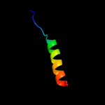
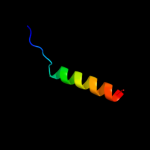
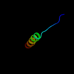
| 1 |
|
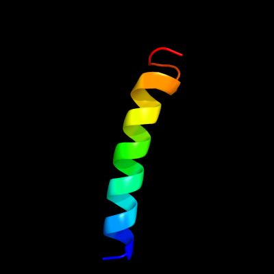
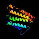
PDB 1yew chain I
Region: 37 - 63
Aligned: 24
Modelled: 27
Confidence: 11.9%
Identity: 13%
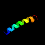
PDB header:oxidoreductase, membrane protein
Chain: I: PDB Molecule:particulate methane monooxygenase, b subunit;
PDBTitle: crystal structure of particulate methane monooxygenase
Phyre2
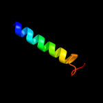
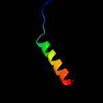
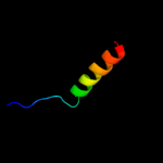
| 2 |
|
PDB 3rgb chain A
Region: 37 - 63
Aligned: 24
Modelled: 27
Confidence: 11.9%
Identity: 13%
PDB header:oxidoreductase
Chain: A: PDB Molecule:methane monooxygenase subunit b2;
PDBTitle: crystal structure of particulate methane monooxygenase from2 methylococcus capsulatus (bath)
Phyre2
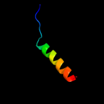
| 3 |
|
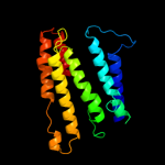
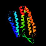
PDB 1xio chain A
Region: 4 - 181
Aligned: 157
Modelled: 178
Confidence: 8.9%
Identity: 17%
PDB header:signaling protein
Chain: A: PDB Molecule:anabaena sensory rhodopsin;
PDBTitle: anabaena sensory rhodopsin
Phyre2
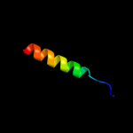
| 4 |
|
PDB 1xio chain A
Region: 4 - 181
Aligned: 157
Modelled: 178
Confidence: 8.9%
Identity: 17%
Fold: Family A G protein-coupled receptor-like
Superfamily: Family A G protein-coupled receptor-like
Family: Bacteriorhodopsin-like
Phyre2
| 5 |
|
PDB 1uaz chain A
Region: 5 - 180
Aligned: 164
Modelled: 176
Confidence: 7.0%
Identity: 10%
Fold: Family A G protein-coupled receptor-like
Superfamily: Family A G protein-coupled receptor-like
Family: Bacteriorhodopsin-like
Phyre2
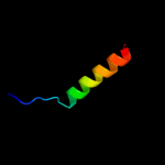
| 6 |
|
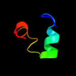
PDB 1dxs chain A
Region: 89 - 114
Aligned: 19
Modelled: 26
Confidence: 6.8%
Identity: 21%
Fold: SAM domain-like
Superfamily: SAM/Pointed domain
Family: SAM (sterile alpha motif) domain
Phyre2
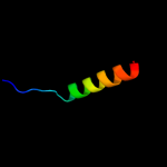
| 7 |
|
PDB 3prq chain L
Region: 105 - 129
Aligned: 25
Modelled: 25
Confidence: 6.3%
Identity: 32%
PDB header:photosynthesis
Chain: L: PDB Molecule:photosystem ii reaction center protein l;
PDBTitle: crystal structure of cyanobacterial photosystem ii in complex with2 terbutryn (part 1 of 2). this file contains first monomer of psii3 dimer
Phyre2
| 8 |
|
PDB 3bz1 chain L
Region: 105 - 129
Aligned: 25
Modelled: 25
Confidence: 6.3%
Identity: 32%
PDB header:electron transport
Chain: L: PDB Molecule:photosystem ii reaction center protein l;
PDBTitle: crystal structure of cyanobacterial photosystem ii (part 12 of 2). this file contains first monomer of psii dimer
Phyre2
| 9 |
|
PDB 3prr chain L
Region: 105 - 129
Aligned: 25
Modelled: 25
Confidence: 6.3%
Identity: 32%
PDB header:photosynthesis
Chain: L: PDB Molecule:photosystem ii reaction center protein l;
PDBTitle: crystal structure of cyanobacterial photosystem ii in complex with2 terbutryn (part 2 of 2). this file contains second monomer of psii3 dimer
Phyre2
| 10 |
|
PDB 1s5l chain L
Region: 105 - 129
Aligned: 25
Modelled: 25
Confidence: 6.3%
Identity: 32%
PDB header:photosynthesis
Chain: L: PDB Molecule:photosystem ii reaction center l protein;
PDBTitle: architecture of the photosynthetic oxygen evolving center
Phyre2
| 11 |
|
PDB 2axt chain L
Region: 105 - 129
Aligned: 25
Modelled: 25
Confidence: 6.3%
Identity: 32%
PDB header:electron transport
Chain: L: PDB Molecule:photosystem ii reaction center l protein;
PDBTitle: crystal structure of photosystem ii from thermosynechococcus elongatus
Phyre2
| 12 |
|
PDB 3arc chain L
Region: 105 - 129
Aligned: 25
Modelled: 25
Confidence: 6.3%
Identity: 32%
PDB header:electron transport, photosynthesis
Chain: L: PDB Molecule:photosystem ii reaction center protein l;
PDBTitle: crystal structure of oxygen-evolving photosystem ii at 1.9 angstrom2 resolution
Phyre2
| 13 |
|
PDB 1s5l chain L
Region: 105 - 129
Aligned: 25
Modelled: 25
Confidence: 6.3%
Identity: 32%
PDB header:photosynthesis
Chain: L: PDB Molecule:photosystem ii reaction center l protein;
PDBTitle: architecture of the photosynthetic oxygen evolving center
Phyre2
| 14 |
|
PDB 3bz2 chain L
Region: 105 - 129
Aligned: 25
Modelled: 25
Confidence: 6.3%
Identity: 32%
PDB header:electron transport
Chain: L: PDB Molecule:photosystem ii reaction center protein l;
PDBTitle: crystal structure of cyanobacterial photosystem ii (part 22 of 2). this file contains second monomer of psii dimer
Phyre2
| 15 |
|
PDB 2axt chain L
Region: 105 - 129
Aligned: 25
Modelled: 25
Confidence: 6.3%
Identity: 32%
PDB header:electron transport
Chain: L: PDB Molecule:photosystem ii reaction center l protein;
PDBTitle: crystal structure of photosystem ii from thermosynechococcus elongatus
Phyre2
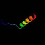
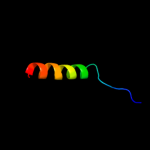
| 16 |
|
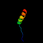
PDB 2axt chain L domain 1
Region: 105 - 129
Aligned: 25
Modelled: 25
Confidence: 6.3%
Identity: 32%
Fold: Single transmembrane helix
Superfamily: Photosystem II reaction center protein L, PsbL
Family: PsbL-like
Phyre2
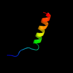
| 17 |
|
PDB 3kzi chain L
Region: 105 - 129
Aligned: 25
Modelled: 25
Confidence: 6.3%
Identity: 32%
PDB header:electron transport
Chain: L: PDB Molecule:photosystem ii reaction center protein l;
PDBTitle: crystal structure of monomeric form of cyanobacterial photosystem ii
Phyre2
| 18 |
|
PDB 3a0h chain L
Region: 105 - 129
Aligned: 25
Modelled: 25
Confidence: 6.2%
Identity: 32%
PDB header:electron transport
Chain: L: PDB Molecule:photosystem ii reaction center protein l;
PDBTitle: crystal structure of i-substituted photosystem ii complex
Phyre2
| 19 |
|
PDB 3a0h chain L
Region: 105 - 129
Aligned: 25
Modelled: 25
Confidence: 6.2%
Identity: 32%
PDB header:electron transport
Chain: L: PDB Molecule:photosystem ii reaction center protein l;
PDBTitle: crystal structure of i-substituted photosystem ii complex
Phyre2
| 20 |
|
PDB 3a0b chain L
Region: 105 - 129
Aligned: 25
Modelled: 25
Confidence: 6.2%
Identity: 32%
PDB header:electron transport
Chain: L: PDB Molecule:photosystem ii reaction center protein l;
PDBTitle: crystal structure of br-substituted photosystem ii complex
Phyre2
| 21 |
|
| 22 |
|