| Secondary structure and disorder prediction | |
| | |
1 | . | . | . | . | . | . | . | . | 10 | . | . | . | . | . | . | . | . | . | 20 | . | . | . | . | . | . | . | . | . | 30 | . | . | . | . | . | . | . | . | . | 40 | . |
| Sequence | |
M | N | R | L | I | E | L | T | G | W | I | V | L | V | V | S | V | I | L | L | G | V | A | S | H | I | D | N | Y | Q | P | P | E | Q | S | A | S | V | Q | H | K |
| Secondary structure | |
|  | | | | | | | | | | | | | | | | | | | | | | | | |
|
|
|
|
|
| | | | | | |
|
|
|
| SS confidence | |
|
|
|
|
|
|
|
|
|
|
|
|
|
|
|
|
|
|
|
|
|
|
|
|
|
|
|
|
|
|
|
|
|
|
|
|
|
|
|
|
|
| Disorder | |
? | ? | ? | ? |
|
|
|
|
|
|
|
|
|
|
|
|
|
|
|
|
|
|
|
|
|
| ? |
|
| ? |
| ? | ? | ? | ? | ? | ? | ? | ? | ? | ? |
| Disorder confidence | |
|
|
|
|
|
|
|
|
|
|
|
|
|
|
|
|
|
|
|
|
|
|
|
|
|
|
|
|
|
|
|
|
|
|
|
|
|
|
|
|
|
| |
| Confidence Key |
| High(9) | |
|
|
|
|
|
|
|
|
|
Low (0) |
| ? | Disordered |
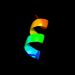
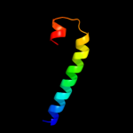
| Alpha helix |
  | Beta strand |
Hover over an aligned region to see model and summary info
Please note, only up to the top 20 hits are modelled to reduce computer load
|
| 1 |
|
PDB 1sr3 chain A
Region: 22 - 38
Aligned: 17
Modelled: 17
Confidence: 14.9%
Identity: 41%
Fold: OB-fold
Superfamily: Heme chaperone CcmE
Family: Heme chaperone CcmE
Phyre2
| 2 |
|
PDB 3o0r chain C
Region: 1 - 25
Aligned: 25
Modelled: 25
Confidence: 12.8%
Identity: 20%
PDB header:immune system/oxidoreductase
Chain: C: PDB Molecule:nitric oxide reductase subunit c;
PDBTitle: crystal structure of nitric oxide reductase from pseudomonas2 aeruginosa in complex with antibody fragment
Phyre2
| 3 |
|
PDB 2apl chain A domain 1
Region: 5 - 15
Aligned: 11
Modelled: 11
Confidence: 9.5%
Identity: 64%
Fold: PG0816-like
Superfamily: PG0816-like
Family: PG0816-like
Phyre2
| 4 |
|
PDB 3ke2 chain A
Region: 3 - 10
Aligned: 8
Modelled: 7
Confidence: 7.7%
Identity: 50%
PDB header:unknown function
Chain: A: PDB Molecule:uncharacterized protein yp_928783.1;
PDBTitle: crystal structure of a duf2131 family protein (sama_2911) from2 shewanella amazonensis sb2b at 2.50 a resolution
Phyre2
| 5 |
|
PDB 1h6g chain A domain 2
Region: 4 - 38
Aligned: 35
Modelled: 35
Confidence: 5.4%
Identity: 29%
Fold: Four-helical up-and-down bundle
Superfamily: alpha-catenin/vinculin-like
Family: alpha-catenin/vinculin
Phyre2
|
| Detailed template information | |
Due to computational demand, binding site predictions are not run for batch jobs
If you want to predict binding sites, please manually submit your model of choice to 3DLigandSite
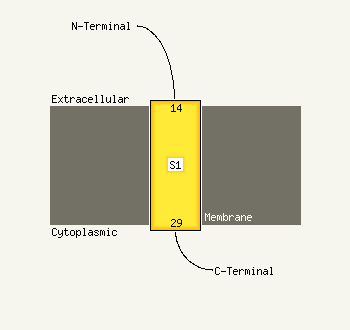
| Transmembrane helix prediction | |
Transmembrane helices have been predicted in your sequence to adopt the topology shown below
Phyre is for academic use only
| Please cite: Protein structure prediction on
the web: a case study using the Phyre server |
| Kelley LA and Sternberg MJE. Nature Protocols
4, 363 - 371 (2009) [pdf] [Import into BibTeX] |
| |
| If you use the binding site
predictions from 3DLigandSite, please also cite: |
| 3DLigandSite: predicting ligand-binding sites using similar structures. |
| Wass MN, Kelley LA and Sternberg
MJ Nucleic Acids Research 38, W469-73 (2010) [PubMed] |
| |
|
|
|
|