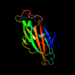
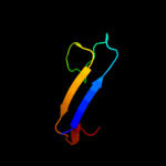
| 1 |
|
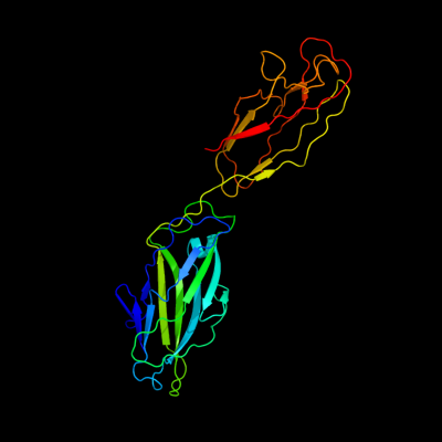
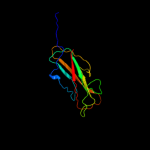
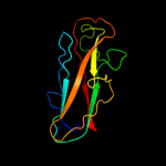
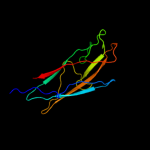
PDB 1klf chain P
Region: 25 - 304
Aligned: 277
Modelled: 280
Confidence: 100.0%
Identity: 50%
PDB header:chaperone/adhesin complex
Chain: P: PDB Molecule:fimh protein;
PDBTitle: fimh adhesin-fimc chaperone complex with d-mannose
Phyre2
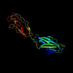
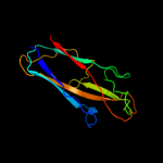
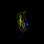
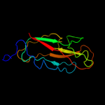
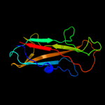
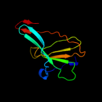
| 2 |
|
PDB 1ze3 chain H domain 1
Region: 183 - 304
Aligned: 121
Modelled: 122
Confidence: 100.0%
Identity: 55%
Fold: Common fold of diphtheria toxin/transcription factors/cytochrome f
Superfamily: Bacterial adhesins
Family: Pilus subunits
Phyre2
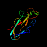
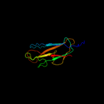
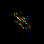
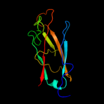
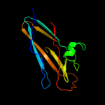
| 3 |
|
PDB 1uwf chain A domain 1
Region: 25 - 182
Aligned: 156
Modelled: 158
Confidence: 99.9%
Identity: 46%
Fold: Common fold of diphtheria toxin/transcription factors/cytochrome f
Superfamily: Bacterial adhesins
Family: Pilus subunits
Phyre2
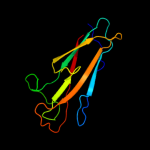
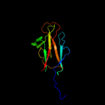
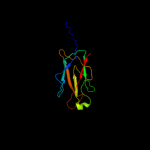
| 4 |
|
PDB 3bwu chain F
Region: 183 - 304
Aligned: 120
Modelled: 122
Confidence: 99.9%
Identity: 16%
PDB header:chaperone, structural, membrane protein
Chain: F: PDB Molecule:protein fimf;
PDBTitle: crystal structure of the ternary complex of fimd (n-terminal domain,2 fimdn) with fimc and the n-terminally truncated pilus subunit fimf3 (fimft)
Phyre2
| 5 |
|
PDB 3jwn chain K
Region: 172 - 304
Aligned: 132
Modelled: 133
Confidence: 99.9%
Identity: 19%
PDB header:protein binding/cell adhesion
Chain: K: PDB Molecule:protein fimf;
PDBTitle: complex of fimc, fimf, fimg and fimh
Phyre2
| 6 |
|
PDB 3bfw chain A
Region: 182 - 303
Aligned: 118
Modelled: 122
Confidence: 99.9%
Identity: 26%
PDB header:structural protein/structural protein
Chain: A: PDB Molecule:protein fimg;
PDBTitle: crystal structure of truncated fimg (fimgt) in complex with the donor2 strand peptide of fimf (dsf)
Phyre2
| 7 |
|
PDB 3jwn chain L
Region: 172 - 304
Aligned: 132
Modelled: 133
Confidence: 99.9%
Identity: 19%
PDB header:protein binding/cell adhesion
Chain: L: PDB Molecule:protein fimf;
PDBTitle: complex of fimc, fimf, fimg and fimh
Phyre2
| 8 |
|
PDB 3jwn chain F
Region: 172 - 304
Aligned: 132
Modelled: 133
Confidence: 99.9%
Identity: 19%
PDB header:protein binding/cell adhesion
Chain: F: PDB Molecule:protein fimf;
PDBTitle: complex of fimc, fimf, fimg and fimh
Phyre2
| 9 |
|
PDB 3jwn chain E
Region: 172 - 304
Aligned: 132
Modelled: 133
Confidence: 99.9%
Identity: 17%
PDB header:protein binding/cell adhesion
Chain: E: PDB Molecule:protein fimf;
PDBTitle: complex of fimc, fimf, fimg and fimh
Phyre2
| 10 |
|
PDB 2j2z chain B domain 1
Region: 171 - 304
Aligned: 129
Modelled: 134
Confidence: 99.9%
Identity: 17%
Fold: Common fold of diphtheria toxin/transcription factors/cytochrome f
Superfamily: Bacterial adhesins
Family: Pilus subunits
Phyre2
| 11 |
|
PDB 2jty chain A
Region: 172 - 304
Aligned: 132
Modelled: 133
Confidence: 99.8%
Identity: 24%
PDB header:structural protein
Chain: A: PDB Molecule:type-1 fimbrial protein, a chain;
PDBTitle: self-complemented variant of fima, the main subunit of type 1 pilus
Phyre2
| 12 |
|
PDB 2jmr chain A
Region: 172 - 304
Aligned: 132
Modelled: 133
Confidence: 99.8%
Identity: 17%
PDB header:cell adhesion
Chain: A: PDB Molecule:fimf;
PDBTitle: nmr structure of the e. coli type 1 pilus subunit fimf
Phyre2
| 13 |
|
PDB 1pdk chain B
Region: 180 - 304
Aligned: 122
Modelled: 125
Confidence: 99.8%
Identity: 19%
Fold: Common fold of diphtheria toxin/transcription factors/cytochrome f
Superfamily: Bacterial adhesins
Family: Pilus subunits
Phyre2
| 14 |
|
PDB 2uy6 chain B domain 1
Region: 171 - 304
Aligned: 129
Modelled: 134
Confidence: 99.8%
Identity: 14%
Fold: Common fold of diphtheria toxin/transcription factors/cytochrome f
Superfamily: Bacterial adhesins
Family: Pilus subunits
Phyre2
| 15 |
|
PDB 2w07 chain B
Region: 180 - 304
Aligned: 109
Modelled: 125
Confidence: 99.7%
Identity: 17%
PDB header:cell adhesion
Chain: B: PDB Molecule:minor pilin subunit papf;
PDBTitle: structural determinants of polymerization reactivity of the2 p pilus adaptor subunit papf
Phyre2
| 16 |
|
PDB 1n12 chain A
Region: 183 - 303
Aligned: 118
Modelled: 121
Confidence: 99.4%
Identity: 19%
Fold: Common fold of diphtheria toxin/transcription factors/cytochrome f
Superfamily: Bacterial adhesins
Family: Pilus subunits
Phyre2
| 17 |
|
PDB 2wmp chain B
Region: 182 - 303
Aligned: 111
Modelled: 122
Confidence: 87.6%
Identity: 19%
PDB header:chaperone
Chain: B: PDB Molecule:papg protein;
PDBTitle: structure of the e. coli chaperone papd in complex with the pilin2 domain of the papgii adhesin
Phyre2
| 18 |
|
PDB 2jna chain A domain 1
Region: 1 - 25
Aligned: 22
Modelled: 25
Confidence: 25.3%
Identity: 23%
Fold: Dodecin subunit-like
Superfamily: YdgH-like
Family: YdgH-like
Phyre2
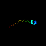
| 19 |
|
PDB 2oz4 chain A domain 1
Region: 206 - 245
Aligned: 40
Modelled: 40
Confidence: 9.7%
Identity: 13%
Fold: Immunoglobulin-like beta-sandwich
Superfamily: Immunoglobulin
Family: C2 set domains
Phyre2
| 20 |
|
PDB 1na8 chain A
Region: 186 - 289
Aligned: 104
Modelled: 104
Confidence: 8.0%
Identity: 18%
Fold: Immunoglobulin-like beta-sandwich
Superfamily: Clathrin adaptor appendage domain
Family: gamma-adaptin C-terminal appendage domain-like
Phyre2
| 21 |
|
| 22 |
|
| 23 |
|
| 24 |
|