| Secondary structure and disorder prediction | |
| | |
1 | . | . | . | . | . | . | . | . | 10 | . | . | . | . | . | . | . | . | . | 20 | . | . | . | . | . | . | . | . | . | 30 | . | . | . | . | . | . | . | . | . | 40 | . | . | . | . | . | . | . | . | . | 50 | . | . | . | . | . | . | . | . | . | 60 |
| Sequence | |
M | K | K | I | L | E | A | I | L | A | I | N | L | A | V | L | S | C | I | V | F | I | N | I | I | L | R | Y | G | F | Q | T | S | I | L | S | V | D | E | L | S | R | Y | L | F | V | W | L | T | F | I | G | A | I | V | A | F | M | D | N |
| Secondary structure | |
|  | | | | | | | | | | | | | | | | | | | | | | | | | | | | |
|
|
|
|
|
| | | | | | | | | | | | | | | | | | | | | | | |
|
| SS confidence | |
|
|
|
|
|
|
|
|
|
|
|
|
|
|
|
|
|
|
|
|
|
|
|
|
|
|
|
|
|
|
|
|
|
|
|
|
|
|
|
|
|
|
|
|
|
|
|
|
|
|
|
|
|
|
|
|
|
|
|
|
| Disorder | |
? |
|
|
|
|
|
|
|
|
|
|
|
|
|
|
|
|
|
|
|
|
|
|
|
|
|
|
|
|
|
|
|
|
|
|
|
|
|
|
|
|
|
|
|
|
|
|
|
|
|
|
|
|
|
|
|
|
|
|
|
| Disorder confidence | |
|
|
|
|
|
|
|
|
|
|
|
|
|
|
|
|
|
|
|
|
|
|
|
|
|
|
|
|
|
|
|
|
|
|
|
|
|
|
|
|
|
|
|
|
|
|
|
|
|
|
|
|
|
|
|
|
|
|
|
|
| |
| | |
. | . | . | . | . | . | . | . | . | 70 | . | . | . | . | . | . | . | . | . | 80 | . | . | . | . | . | . | . | . | . | 90 | . | . | . | . | . | . | . | . | . | 100 | . | . | . | . | . | . | . | . | . | 110 | . | . | . | . | . | . | . | . | . | 120 |
| Sequence | |
A | H | V | Q | V | T | F | L | V | E | K | L | S | P | A | W | Q | R | R | V | A | L | V | T | H | S | L | I | L | F | I | C | G | A | L | A | W | G | A | T | L | K | T | I | Q | D | W | S | D | Y | S | P | I | L | G | L | P | I | G | L |
| Secondary structure | |
| | | | | | | | | | |
|
| | | | | | | | | | | | | | | | | | | | | | | | | | | | | | | | | | |
|
|
|
|
| | |
|
|
| | | |
| SS confidence | |
|
|
|
|
|
|
|
|
|
|
|
|
|
|
|
|
|
|
|
|
|
|
|
|
|
|
|
|
|
|
|
|
|
|
|
|
|
|
|
|
|
|
|
|
|
|
|
|
|
|
|
|
|
|
|
|
|
|
|
|
| Disorder | |
|
|
|
|
|
|
|
|
|
|
|
|
|
|
|
|
|
|
|
|
|
|
|
|
|
|
|
|
|
|
|
|
|
|
|
|
|
|
|
|
|
|
|
|
|
| ? | ? | ? | ? | ? | ? | ? |
| ? | ? | ? |
|
|
|
| Disorder confidence | |
|
|
|
|
|
|
|
|
|
|
|
|
|
|
|
|
|
|
|
|
|
|
|
|
|
|
|
|
|
|
|
|
|
|
|
|
|
|
|
|
|
|
|
|
|
|
|
|
|
|
|
|
|
|
|
|
|
|
|
|
| |
| | |
. | . | . | . | . | . | . | . | . | 130 | . | . | . | . | . | . | . | . | . | 140 | . | . | . | . | . | . | . | . | . | 150 | . | . | . | . | . | . | . |
| Sequence | |
M | Y | A | A | C | L | P | T | S | L | V | I | A | F | F | E | L | R | H | L | Y | Q | L | I | T | R | S | N | S | L | T | S | P | P | Q | G | A |
| Secondary structure | |
| | | | | | | | | | | | | | | | | | | | | | | | |
|
|
|
|
|
|
|
|
|
|
|
|
| SS confidence | |
|
|
|
|
|
|
|
|
|
|
|
|
|
|
|
|
|
|
|
|
|
|
|
|
|
|
|
|
|
|
|
|
|
|
|
|
|
| Disorder | |
|
|
|
|
|
|
|
|
|
|
|
|
|
|
|
|
|
|
|
|
|
|
|
|
|
|
| ? | ? | ? | ? | ? | ? | ? | ? | ? | ? |
| Disorder confidence | |
|
|
|
|
|
|
|
|
|
|
|
|
|
|
|
|
|
|
|
|
|
|
|
|
|
|
|
|
|
|
|
|
|
|
|
|
|
| |
| Confidence Key |
| High(9) | |
|
|
|
|
|
|
|
|
|
Low (0) |
| ? | Disordered |
| Alpha helix |
  | Beta strand |
Hover over an aligned region to see model and summary info
Please note, only up to the top 20 hits are modelled to reduce computer load
|
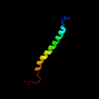
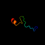
| 1 |
|
PDB 2knc chain A
Region: 108 - 157
Aligned: 50
Modelled: 50
Confidence: 47.7%
Identity: 14%
PDB header:cell adhesion
Chain: A: PDB Molecule:integrin alpha-iib;
PDBTitle: platelet integrin alfaiib-beta3 transmembrane-cytoplasmic2 heterocomplex
Phyre2
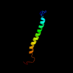
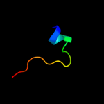
| 2 |
|
PDB 1yle chain A domain 1
Region: 21 - 80
Aligned: 41
Modelled: 40
Confidence: 22.7%
Identity: 27%
Fold: Acyl-CoA N-acyltransferases (Nat)
Superfamily: Acyl-CoA N-acyltransferases (Nat)
Family: AstA-like
Phyre2
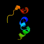
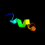
| 3 |
|
PDB 2k42 chain A
Region: 52 - 84
Aligned: 33
Modelled: 33
Confidence: 8.5%
Identity: 18%
PDB header:signaling protein
Chain: A: PDB Molecule:wiskott-aldrich syndrome protein;
PDBTitle: solution structure of the gtpase binding domain of wasp in2 complex with espfu, an ehec effector
Phyre2
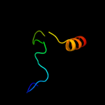
| 4 |
|
PDB 2odb chain B
Region: 52 - 81
Aligned: 30
Modelled: 30
Confidence: 7.3%
Identity: 23%
PDB header:protein binding
Chain: B: PDB Molecule:serine/threonine-protein kinase pak 6;
PDBTitle: the crystal structure of human cdc42 in complex with the crib domain2 of human p21-activated kinase 6 (pak6)
Phyre2
| 5 |
|
PDB 3k6g chain A
Region: 22 - 39
Aligned: 17
Modelled: 18
Confidence: 6.7%
Identity: 0%
PDB header:protein binding
Chain: A: PDB Molecule:telomeric repeat-binding factor 2-interacting protein 1;
PDBTitle: crystal structure of rap1 and trf2 complex
Phyre2
| 6 |
|
PDB 2kpa chain A
Region: 63 - 79
Aligned: 17
Modelled: 17
Confidence: 6.1%
Identity: 29%
PDB header:hydrolase
Chain: A: PDB Molecule:arno(375-400);
PDBTitle: specific motifs of the v-atpase a2-subunit isoform interact2 with catalytic and regulatory domains of arno
Phyre2
|
| Detailed template information | |
Due to computational demand, binding site predictions are not run for batch jobs
If you want to predict binding sites, please manually submit your model of choice to 3DLigandSite
Phyre is for academic use only
| Please cite: Protein structure prediction on
the web: a case study using the Phyre server |
| Kelley LA and Sternberg MJE. Nature Protocols
4, 363 - 371 (2009) [pdf] [Import into BibTeX] |
| |
| If you use the binding site
predictions from 3DLigandSite, please also cite: |
| 3DLigandSite: predicting ligand-binding sites using similar structures. |
| Wass MN, Kelley LA and Sternberg
MJ Nucleic Acids Research 38, W469-73 (2010) [PubMed] |
| |
|
|
|
|