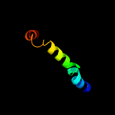
Phyre Home Retrieve Phyre Job Id
Emaill.a.kelley@imperial.ac.uk
DescriptionQ05807
DateThu Jan 5 12:33:22 GMT 2012
Unique Job ID2e580a9590512940

Summary 

Top model
Image coloured by rainbow N → C terminus
Model (left) based on template c3rkoF_
Top template information
PDB header:oxidoreductase
Chain: F: PDB Molecule:nadh-quinone oxidoreductase subunit j;
PDBTitle: crystal structure of the membrane domain of respiratory complex i from2 e. coli at 3.0 angstrom resolution
Confidence and coverage
Confidence: 21.7% Coverage: 49%
42 residues ( 49% of your sequence) have been modelled with 21.7% confidence by the single highest scoring template.
You may wish to submit your sequence to Phyrealarm. This will automatically scan your sequence every week for new potential templates as they appear in the Phyre2 library.
Please note: You must be registered and logged in to use Phyrealarm.
3D viewing
Interactive 3D view in Jmol

Sequence analysis 

Secondary structure and disorder prediction 

   1........10.........20.........30.........40.........50.........60
Sequence  MMKVIFTSNRFIDFLIRLLITAIVISPVIIWSWDTVKETTADGMLAAAFVILYSGVLLFI
Secondary structure 


SS confidence 


Disorder  ???

??


????


Disorder confidence 


   .........70.........80......
Sequence  LYFCFSALTDLQKPDERKSDERNEDE
Secondary structure 


SS confidence 


Disorder 


??????????????
Disorder confidence 


Confidence Key
High(9)                    Low (0)
?Disordered
Alpha helix
Beta strand

Domain analysis 

Hover over an aligned region to see model and summary info

Please note, only up to the top 20 hits are modelled to reduce computer load

RankAligned region

PDB 3rko chain F

3D model

Region: 41 - 83
Aligned: 42
Modelled: 43
Confidence: 21.7%
Identity: 14%
PDB header:oxidoreductase
Chain: F: PDB Molecule:nadh-quinone oxidoreductase subunit j;
PDBTitle: crystal structure of the membrane domain of respiratory complex i from2 e. coli at 3.0 angstrom resolution

Phyre2

PDB 1utc chain A domain 2

3D model

Region: 27 - 33
Aligned: 7
Modelled: 7
Confidence: 18.2%
Identity: 43%
Fold: 7-bladed beta-propeller
Superfamily: Clathrin heavy-chain terminal domain
Family: Clathrin heavy-chain terminal domain

Phyre2

PDB 1bpo chain A

3D model

Region: 27 - 33
Aligned: 7
Modelled: 7
Confidence: 17.9%
Identity: 43%
PDB header:membrane protein
Chain: A: PDB Molecule:protein (clathrin);
PDBTitle: clathrin heavy-chain terminal domain and linker

Phyre2

PDB 1c9l chain A

3D model

Region: 27 - 33
Aligned: 7
Modelled: 7
Confidence: 17.3%
Identity: 43%
PDB header:endocytosis/exocytosis
Chain: A: PDB Molecule:clathrin;
PDBTitle: peptide-in-groove interactions link target proteins to the2 b-propeller of clathrin

Phyre2

PDB 3qnq chain D

3D model

Region: 40 - 81
Aligned: 42
Modelled: 42
Confidence: 16.7%
Identity: 5%
PDB header:membrane protein, transport protein
Chain: D: PDB Molecule:pts system, cellobiose-specific iic component;
PDBTitle: crystal structure of the transporter chbc, the iic component from the2 n,n'-diacetylchitobiose-specific phosphotransferase system

Phyre2

PDB 1xi4 chain D

3D model

Region: 27 - 33
Aligned: 7
Modelled: 7
Confidence: 13.7%
Identity: 43%
PDB header:endocytosis/exocytosis
Chain: D: PDB Molecule:clathrin heavy chain;
PDBTitle: clathrin d6 coat

Phyre2

PDB 2q04 chain C

3D model

Region: 23 - 34
Aligned: 12
Modelled: 12
Confidence: 10.8%
Identity: 25%
PDB header:transferase
Chain: C: PDB Molecule:acetoin utilization protein;
PDBTitle: crystal structure of acetoin utilization protein (zp_00540088.1) from2 exiguobacterium sibiricum 255-15 at 2.33 a resolution

Phyre2

PDB 2k59 chain B

3D model

Region: 12 - 29
Aligned: 18
Modelled: 18
Confidence: 6.6%
Identity: 28%
PDB header:transport protein
Chain: B: PDB Molecule:neuronal acetylcholine receptor subunit beta-2;
PDBTitle: nmr structures of the second transmembrane domain of the2 neuronal acetylcholine receptor beta 2 subunit

Phyre2

PDB 3c8e chain B

3D model

Region: 27 - 41
Aligned: 15
Modelled: 15
Confidence: 5.9%
Identity: 20%
PDB header:transferase
Chain: B: PDB Molecule:yghu, glutathione s-transferase homologue;
PDBTitle: crystal structure analysis of yghu from e. coli

Phyre2

PDB 1qhk chain A

3D model

Region: 26 - 37
Aligned: 12
Modelled: 12
Confidence: 5.8%
Identity: 25%
Fold: MbtH/L9 domain-like
Superfamily: L9 N-domain-like
Family: N-terminal domain of RNase HI

Phyre2

PDB 3bsu chain F

3D model

Region: 26 - 37
Aligned: 12
Modelled: 12
Confidence: 5.6%
Identity: 8%
PDB header:hydrolase/rna/dna
Chain: F: PDB Molecule:ribonuclease h1;
PDBTitle: hybrid-binding domain of human rnase h1 in complex with 12-2 mer rna/dna

Phyre2
1

c3rkoF_
2

d1utca2
3

c1bpoA_
4

c1c9lA_
5

c3qnqD_
6

c1xi4D_
7

c2q04C_
8

c2k59B_
9

c3c8eB_
10

d1qhka_
11

c3bsuF_


Detailed template information 

#
Template Alignment Coverage3D Model Confidence
% i.d. Template Information
1c3rkoF_


21.7 14 PDB header:oxidoreductase
Chain: F: PDB Molecule:nadh-quinone oxidoreductase subunit j;
PDBTitle: crystal structure of the membrane domain of respiratory complex i from2 e. coli at 3.0 angstrom resolution
2d1utca2


18.2 43 Fold:7-bladed beta-propeller
Superfamily:Clathrin heavy-chain terminal domain
Family:Clathrin heavy-chain terminal domain
3c1bpoA_


17.9 43 PDB header:membrane protein
Chain: A: PDB Molecule:protein (clathrin);
PDBTitle: clathrin heavy-chain terminal domain and linker
4c1c9lA_


17.3 43 PDB header:endocytosis/exocytosis
Chain: A: PDB Molecule:clathrin;
PDBTitle: peptide-in-groove interactions link target proteins to the2 b-propeller of clathrin
5c3qnqD_


16.7 5 PDB header:membrane protein, transport protein
Chain: D: PDB Molecule:pts system, cellobiose-specific iic component;
PDBTitle: crystal structure of the transporter chbc, the iic component from the2 n,n'-diacetylchitobiose-specific phosphotransferase system
6c1xi4D_


13.7 43 PDB header:endocytosis/exocytosis
Chain: D: PDB Molecule:clathrin heavy chain;
PDBTitle: clathrin d6 coat
7c2q04C_


10.8 25 PDB header:transferase
Chain: C: PDB Molecule:acetoin utilization protein;
PDBTitle: crystal structure of acetoin utilization protein (zp_00540088.1) from2 exiguobacterium sibiricum 255-15 at 2.33 a resolution
8c2k59B_


6.6 28 PDB header:transport protein
Chain: B: PDB Molecule:neuronal acetylcholine receptor subunit beta-2;
PDBTitle: nmr structures of the second transmembrane domain of the2 neuronal acetylcholine receptor beta 2 subunit
9c3c8eB_


5.9 20 PDB header:transferase
Chain: B: PDB Molecule:yghu, glutathione s-transferase homologue;
PDBTitle: crystal structure analysis of yghu from e. coli
10d1qhka_


5.8 25 Fold:MbtH/L9 domain-like
Superfamily:L9 N-domain-like
Family:N-terminal domain of RNase HI
11c3bsuF_


5.6 8 PDB header:hydrolase/rna/dna
Chain: F: PDB Molecule:ribonuclease h1;
PDBTitle: hybrid-binding domain of human rnase h1 in complex with 12-2 mer rna/dna

Binding site prediction 

Due to computational demand, binding site predictions are not run for batch jobs

If you want to predict binding sites, please manually submit your model of choice to 3DLigandSite


Phyre is for academic use only

Please cite: Protein structure prediction on the web: a case study using the Phyre server
Kelley LA and Sternberg MJE. Nature Protocols 4, 363 - 371 (2009) [pdf] [Import into BibTeX]
 
If you use the binding site predictions from 3DLigandSite, please also cite:
3DLigandSite: predicting ligand-binding sites using similar structures.
Wass MN, Kelley LA and Sternberg MJ Nucleic Acids Research 38, W469-73 (2010) [PubMed]
 
© Structural Bioinformatics Group
Imperial College London
Lawrence Kelley, Benjamin Jefferys
Disclaimer
Terms and Conditions
Component software
Template detection: HHpred 1.51
Secondary structure prediction: Psi-pred 2.5
Disorder prediction: Disopred 2.4
Transmembrane prediction: Memsat_SVM
Multi-template modelling and ab initio: Poing 1.0