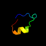
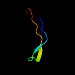
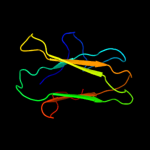
1 c3qsqA_
41.1
31
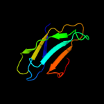
PDB header: viral proteinChain: A: PDB Molecule: capsid polyprotein;PDBTitle: crystal structure of the projection domain of the human astrovirus2 capsid protein
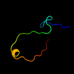
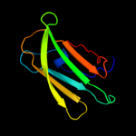
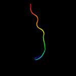
2 c3bdkB_
41.0
17
PDB header: lyaseChain: B: PDB Molecule: d-mannonate dehydratase;PDBTitle: crystal structure of streptococcus suis mannonate2 dehydratase complexed with substrate analogue
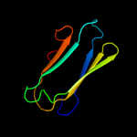
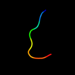
3 d1tz9a_
27.6
22
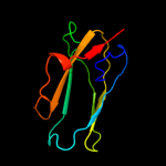
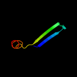
Fold: TIM beta/alpha-barrelSuperfamily: Xylose isomerase-likeFamily: UxuA-like4 c3ba3A_
22.0
22
PDB header: oxidoreductaseChain: A: PDB Molecule: pyridoxamine 5'-phosphate oxidase-like protein;PDBTitle: crystal structure of pyridoxamine 5'-phosphate oxidase-like protein2 (np_783940.1) from lactobacillus plantarum at 1.55 a resolution
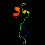
5 c3cnkB_
19.0
14
PDB header: structural proteinChain: B: PDB Molecule: filamin-a;PDBTitle: crystal structure of the dimerization domain of human2 filamin a
6 d2d7ma1
12.9
18
Fold: Immunoglobulin-like beta-sandwichSuperfamily: E set domainsFamily: Filamin repeat (rod domain)7 d2q79a1
12.2
19
Fold: Ferredoxin-likeSuperfamily: Viral DNA-binding domainFamily: Viral DNA-binding domain8 d2bp3a1
11.3
18
Fold: Immunoglobulin-like beta-sandwichSuperfamily: E set domainsFamily: Filamin repeat (rod domain)9 d1gk8a2
10.7
26
Fold: Ferredoxin-likeSuperfamily: RuBisCO, large subunit, small (N-terminal) domainFamily: Ribulose 1,5-bisphosphate carboxylase-oxygenase10 d1rbla2
10.7
21
Fold: Ferredoxin-likeSuperfamily: RuBisCO, large subunit, small (N-terminal) domainFamily: Ribulose 1,5-bisphosphate carboxylase-oxygenase11 c3iswA_
9.6
14
PDB header: structural proteinChain: A: PDB Molecule: filamin-a;PDBTitle: crystal structure of filamin-a immunoglobulin-like repeat 21 bound to2 an n-terminal peptide of cftr
12 d1wf9a1
9.6
36
Fold: beta-Grasp (ubiquitin-like)Superfamily: Ubiquitin-likeFamily: Ubiquitin-related13 c2nytB_
9.0
63
PDB header: hydrolaseChain: B: PDB Molecule: probable c->u-editing enzyme apobec-2;PDBTitle: the apobec2 crystal structure and functional implications2 for aid
14 c1vknC_
9.0
19
PDB header: oxidoreductaseChain: C: PDB Molecule: n-acetyl-gamma-glutamyl-phosphate reductase;PDBTitle: crystal structure of n-acetyl-gamma-glutamyl-phosphate reductase2 (tm1782) from thermotoga maritima at 1.80 a resolution
15 c2kboA_
8.8
67
PDB header: hydrolaseChain: A: PDB Molecule: dna dc->du-editing enzyme apobec-3g;PDBTitle: structure, interaction, and real-time monitoring of the2 enzymatic reaction of wild type apobec3g
16 d2o62a2
8.7
24
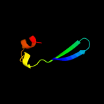
Fold: LipocalinsSuperfamily: LipocalinsFamily: All1756-like17 c2eecA_
8.4
23
PDB header: structural proteinChain: A: PDB Molecule: filamin-b;PDBTitle: solution structure of the 23th filamin domain from human2 filamin-b
18 d8ruca2
7.8
21
Fold: Ferredoxin-likeSuperfamily: RuBisCO, large subunit, small (N-terminal) domainFamily: Ribulose 1,5-bisphosphate carboxylase-oxygenase19 d2e9ia1
7.5
16
Fold: Immunoglobulin-like beta-sandwichSuperfamily: E set domainsFamily: Filamin repeat (rod domain)20 c2zw3B_
7.5
40
PDB header: cell adhesionChain: B: PDB Molecule: gap junction beta-2 protein;PDBTitle: structure of the connexin-26 gap junction channel at 3.52 angstrom resolution
21 c3khpB_
not modelled
7.3
14
PDB header: oxidoreductaseChain: B: PDB Molecule: maoc family protein;PDBTitle: crystal structure of a possible dehydrogenase from2 mycobacterium tuberculosis at 2.3a resolution
22 c1moxB_
not modelled
7.2
14
PDB header: transferase/growth factorChain: B: PDB Molecule: epidermal growth factor receptor;PDBTitle: crystal structure of human epidermal growth factor receptor (residues2 1-501) in complex with tgf-alpha
23 d1zl0a2
not modelled
6.9
54
Fold: Flavodoxin-likeSuperfamily: Class I glutamine amidotransferase-likeFamily: LD-carboxypeptidase A N-terminal domain-like24 c3hsbB_
not modelled
6.7
42
PDB header: rna binding protein/rnaChain: B: PDB Molecule: protein hfq;PDBTitle: crystal structure of ymah (hfq) from bacillus subtilis in complex with2 an rna aptamer
25 d1k8wa3
not modelled
6.6
17
Fold: PUA domain-likeSuperfamily: PUA domain-likeFamily: PUA domain26 c2e9jA_
not modelled
6.3
18
PDB header: structural proteinChain: A: PDB Molecule: filamin-b;PDBTitle: solution structure of the 14th filamin domain from human2 filamin-b
27 d2o62a1
not modelled
6.3
25
Fold: LipocalinsSuperfamily: LipocalinsFamily: All1756-like28 c2jf1A_
not modelled
6.2
18
PDB header: cell adhesionChain: A: PDB Molecule: filamin-a;PDBTitle: crystal structure of the filamin a repeat 21 complexed with2 the integrin beta2 cytoplasmic tail peptide
29 d1ej7l2
not modelled
6.0
29
Fold: Ferredoxin-likeSuperfamily: RuBisCO, large subunit, small (N-terminal) domainFamily: Ribulose 1,5-bisphosphate carboxylase-oxygenase30 c2e2zA_
not modelled
5.9
35
PDB header: protein transport, chaperone regulatorChain: A: PDB Molecule: tim15;PDBTitle: solution nmr structure of yeast tim15, co-chaperone of2 mitochondrial hsp70
31 d1bwva2
not modelled
5.9
26
Fold: Ferredoxin-likeSuperfamily: RuBisCO, large subunit, small (N-terminal) domainFamily: Ribulose 1,5-bisphosphate carboxylase-oxygenase32 d1dbda_
not modelled
5.8
13
Fold: Ferredoxin-likeSuperfamily: Viral DNA-binding domainFamily: Viral DNA-binding domain33 d1hk9a_
not modelled
5.8
39
Fold: Sm-like foldSuperfamily: Sm-like ribonucleoproteinsFamily: Pleiotropic translational regulator Hfq34 d1f9fa_
not modelled
5.7
5
Fold: Ferredoxin-likeSuperfamily: Viral DNA-binding domainFamily: Viral DNA-binding domain35 d1bxna2
not modelled
5.7
24
Fold: Ferredoxin-likeSuperfamily: RuBisCO, large subunit, small (N-terminal) domainFamily: Ribulose 1,5-bisphosphate carboxylase-oxygenase36 d2b59b2
not modelled
5.6
28
Fold: Prealbumin-likeSuperfamily: Carboxypeptidase regulatory domain-likeFamily: Pre-dockerin domain37 c3g23A_
not modelled
5.6
23
PDB header: hydrolaseChain: A: PDB Molecule: ld-carboxypeptidase a;PDBTitle: crystal structure of a ld-carboxypeptidase a (saro_1426) from2 novosphingobium aromaticivorans dsm at 1.89 a resolution
38 c1rcxH_
not modelled
5.6
24
PDB header: lyase (carbon-carbon)Chain: H: PDB Molecule: ribulose bisphosphate carboxylase/oxygenase;PDBTitle: non-activated spinach rubisco in complex with its substrate2 ribulose-1,5-bisphosphate
39 c2q49B_
not modelled
5.4
19
PDB header: oxidoreductaseChain: B: PDB Molecule: probable n-acetyl-gamma-glutamyl-phosphate reductase;PDBTitle: ensemble refinement of the protein crystal structure of gene product2 from arabidopsis thaliana at2g19940
40 d1svda2
not modelled
5.4
24
Fold: Ferredoxin-likeSuperfamily: RuBisCO, large subunit, small (N-terminal) domainFamily: Ribulose 1,5-bisphosphate carboxylase-oxygenase41 d2dtge4
not modelled
5.4
21
Fold: Leucine-rich repeat, LRR (right-handed beta-alpha superhelix)Superfamily: L domain-likeFamily: L domain42 d2auna2
not modelled
5.3
54
Fold: Flavodoxin-likeSuperfamily: Class I glutamine amidotransferase-likeFamily: LD-carboxypeptidase A N-terminal domain-like43 c3b2uI_
not modelled
5.2
14
PDB header: immune system/transferaseChain: I: PDB Molecule: epidermal growth factor receptor;PDBTitle: crystal structure of isolated domain iii of the extracellular region2 of the epidermal growth factor receptor in complex with the fab3 fragment of imc-11f8