Phyre Home Retrieve Phyre Job Id
Emaill.a.kelley@imperial.ac.uk
DescriptionP05041
DateThu Jan 5 10:58:36 GMT 2012
Unique Job ID22e36ecb8efed07a

Summary 

Top model
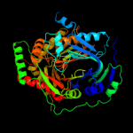
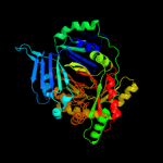
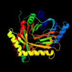
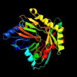
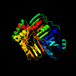
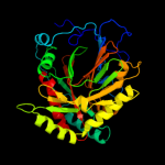
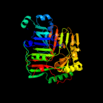
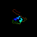
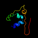
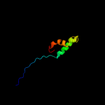
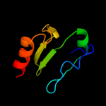
Image coloured by rainbow N → C terminus
Model (left) based on template d1i1qa_
Top template information
Fold:ADC synthase
Superfamily:ADC synthase
Family:ADC synthase
Confidence and coverage
Confidence:100.0% Coverage: 98%
445 residues ( 98% of your sequence) have been modelled with 100.0% confidence by the single highest scoring template.
3D viewing
Interactive 3D view in Jmol

Sequence analysis 

Secondary structure and disorder prediction 

   1........10.........20.........30.........40.........50.........60
Sequence  MKTLSPAVITLLWRQDAAEFYFSRLSHLPWAMLLHSGYADHPYSRFDIVVAEPICTLTTF
Secondary structure 


SS confidence 


Disorder  ??????


?????


Disorder confidence 


   .........70.........80.........90.........100.........110.........120
Sequence  GKETVVSESEKRTTTTDDPLQVLQQVLDRADIRPTHNEDLPFQGGALGLFGYDLGRRFES
Secondary structure 


SS confidence 


Disorder 


????


????
?


Disorder confidence 


   .........130.........140.........150.........160.........170.........180
Sequence  LPEIAEQDIVLPDMAVGIYDWALIVDHQRHTVSLLSHNDVNARRAWLESQQFSPQEDFTL
Secondary structure 


SS confidence 


Disorder 


?


?????????????
Disorder confidence 


   .........190.........200.........210.........220.........230.........240
Sequence  TSDWQSNMTREQYGEKFRQVQEYLHSGDCYQVNLAQRFHATYSGDEWQAFLQLNQANRAP
Secondary structure 


SS confidence 


Disorder  ???


Disorder confidence 


   .........250.........260.........270.........280.........290.........300
Sequence  FSAFLRLEQGAILSLSPERFILCDNSEIQTRPIKGTLPRLPDPQEDSKQAVKLANSAKDR
Secondary structure 


SS confidence 


Disorder 


??


????


Disorder confidence 


   .........310.........320.........330.........340.........350.........360
Sequence  AENLMIVDLMRNDIGRVAVAGSVKVPELFVVEPFPAVHHLVSTITAQLPEQLHASDLLRA
Secondary structure 


SS confidence 


Disorder 


Disorder confidence 


   .........370.........380.........390.........400.........410.........420
Sequence  AFPGGSITGAPKVRAMEIIDELEPQRRNAWCGSIGYLSFCGNMDTSITIRTLTAINGQIF
Secondary structure 


SS confidence 


Disorder 


Disorder confidence 


   .........430.........440.........450...
Sequence  CSAGGGIVADSQEEAEYQETFDKVNRILKQLEK
Secondary structure 


SS confidence 


Disorder 


??
Disorder confidence 


Confidence Key
High(9)                    Low (0)
?Disordered
Alpha helix
Beta strand

Domain analysis 

Hover over an aligned region to see model and summary info

Please note, only up to the top 20 hits are modelled to reduce computer load

RankAligned region

PDB 1i1q chain A

3D model

Region: 2 - 452
Aligned: 445
Modelled: 447
Confidence: 100.0%
Identity: 27%
Fold: ADC synthase
Superfamily: ADC synthase
Family: ADC synthase

Phyre2

PDB 1i7q chain A

3D model

Region: 4 - 452
Aligned: 443
Modelled: 449
Confidence: 100.0%
Identity: 28%
Fold: ADC synthase
Superfamily: ADC synthase
Family: ADC synthase

Phyre2

PDB 1k0g chain A

3D model

Region: 1 - 453
Aligned: 420
Modelled: 429
Confidence: 100.0%
Identity: 100%
Fold: ADC synthase
Superfamily: ADC synthase
Family: ADC synthase

Phyre2

PDB 1qdl chain A

3D model

Region: 8 - 452
Aligned: 410
Modelled: 429
Confidence: 100.0%
Identity: 32%
Fold: ADC synthase
Superfamily: ADC synthase
Family: ADC synthase

Phyre2

PDB 2g5f chain A domain 1

3D model

Region: 9 - 451
Aligned: 422
Modelled: 441
Confidence: 100.0%
Identity: 20%
Fold: ADC synthase
Superfamily: ADC synthase
Family: ADC synthase

Phyre2

PDB 2i6y chain A

3D model

Region: 9 - 451
Aligned: 408
Modelled: 443
Confidence: 100.0%
Identity: 21%
PDB header:lyase
Chain: A: PDB Molecule:anthranilate synthase component i, putative;
PDBTitle: structure and mechanism of mycobacterium tuberculosis salicylate2 synthase, mbti

Phyre2

PDB 2fn0 chain A domain 1

3D model

Region: 8 - 451
Aligned: 400
Modelled: 420
Confidence: 100.0%
Identity: 20%
Fold: ADC synthase
Superfamily: ADC synthase
Family: ADC synthase

Phyre2

PDB 3h9m chain A

3D model

Region: 12 - 452
Aligned: 386
Modelled: 398
Confidence: 100.0%
Identity: 28%
PDB header:lyase
Chain: A: PDB Molecule:p-aminobenzoate synthetase, component i;
PDBTitle: crystal structure of para-aminobenzoate synthetase,2 component i from cytophaga hutchinsonii

Phyre2

PDB 3os6 chain A

3D model

Region: 33 - 451
Aligned: 358
Modelled: 367
Confidence: 100.0%
Identity: 25%
PDB header:isomerase
Chain: A: PDB Molecule:isochorismate synthase dhbc;
PDBTitle: crystal structure of putative 2,3-dihydroxybenzoate-specific2 isochorismate synthase, dhbc from bacillus anthracis.

Phyre2

PDB 3hwo chain B

3D model

Region: 48 - 451
Aligned: 365
Modelled: 367
Confidence: 100.0%
Identity: 22%
PDB header:isomerase
Chain: B: PDB Molecule:isochorismate synthase entc;
PDBTitle: crystal structure of escherichia coli enterobactin-specific2 isochorismate synthase entc in complex with isochorismate

Phyre2

PDB 3bzn chain A domain 1

3D model

Region: 2 - 452
Aligned: 395
Modelled: 409
Confidence: 100.0%
Identity: 19%
Fold: ADC synthase
Superfamily: ADC synthase
Family: ADC synthase

Phyre2

PDB 3r74 chain B

3D model

Region: 96 - 451
Aligned: 351
Modelled: 356
Confidence: 100.0%
Identity: 17%
PDB header:lyase, biosynthetic protein
Chain: B: PDB Molecule:anthranilate/para-aminobenzoate synthases component i;
PDBTitle: crystal structure of 2-amino-2-desoxyisochorismate synthase (adic)2 synthase phze from burkholderia lata 383

Phyre2

PDB 3gse chain A

3D model

Region: 17 - 447
Aligned: 377
Modelled: 399
Confidence: 100.0%
Identity: 22%
PDB header:isomerase
Chain: A: PDB Molecule:menaquinone-specific isochorismate synthase;
PDBTitle: crystal structure of menaquinone-specific isochorismate synthase from2 yersinia pestis co92

Phyre2

PDB 3nqk chain A

3D model

Region: 388 - 424
Aligned: 37
Modelled: 37
Confidence: 35.2%
Identity: 27%
PDB header:structural genomics, unknown function
Chain: A: PDB Molecule:uncharacterized protein;
PDBTitle: crystal structure of a structural genomics, unknown function2 (bacova_03322) from bacteroides ovatus at 2.61 a resolution

Phyre2

PDB 1cvr chain A domain 1

3D model

Region: 390 - 432
Aligned: 38
Modelled: 43
Confidence: 34.2%
Identity: 24%
Fold: Immunoglobulin-like beta-sandwich
Superfamily: E set domains
Family: Gingipain R (RgpB), C-terminal domain

Phyre2

PDB 1wqk chain A

3D model

Region: 384 - 397
Aligned: 13
Modelled: 14
Confidence: 32.5%
Identity: 46%
PDB header:toxin
Chain: A: PDB Molecule:toxin apetx1;
PDBTitle: solution structure of apetx1, a specific peptide inhibitor2 of human ether-a-go-go-related gene potassium channels3 from the venom of the sea anemone anthopleura4 elegantissima: a new fold for an herg toxin

Phyre2

PDB 1ugp chain A

3D model

Region: 285 - 341
Aligned: 43
Modelled: 57
Confidence: 22.1%
Identity: 23%
Fold: Nitrile hydratase alpha chain
Superfamily: Nitrile hydratase alpha chain
Family: Nitrile hydratase alpha chain

Phyre2

PDB 2qdy chain A domain 1

3D model

Region: 285 - 341
Aligned: 43
Modelled: 57
Confidence: 19.7%
Identity: 12%
Fold: Nitrile hydratase alpha chain
Superfamily: Nitrile hydratase alpha chain
Family: Nitrile hydratase alpha chain

Phyre2

PDB 2k27 chain A

3D model

Region: 266 - 323
Aligned: 52
Modelled: 58
Confidence: 16.7%
Identity: 19%
PDB header:transcription regulator
Chain: A: PDB Molecule:paired box protein pax-8;
PDBTitle: solution structure of human pax8 paired box domain

Phyre2

PDB 2npb chain A

3D model

Region: 390 - 452
Aligned: 61
Modelled: 63
Confidence: 14.3%
Identity: 20%
PDB header:oxidoreductase
Chain: A: PDB Molecule:selenoprotein w;
PDBTitle: nmr solution structure of mouse selw

Phyre2
1

d1i1qa_
2

d1i7qa_
3

d1k0ga_
4

d1qdla_
5

d2g5fa1
6

c2i6yA_
7

d2fn0a1
8

c3h9mA_
9

c3os6A_
10

c3hwoB_
11

d3bzna1
12

c3r74B_
13

c3gseA_
14

c3nqkA_
15

d1cvra1
16

c1wqkA_
17

d1ugpa_
18

d2qdya1
19

c2k27A_
20

c2npbA_
21


22


23


24


25


26


27


28


29


30


31


32


33


34


35


36


37


38


39


40


41


42


43


44


45


46


Detailed template information 

#
Template Alignment Coverage3D Model Confidence
% i.d. Template Information
1d1i1qa_


100.0 27 Fold:ADC synthase
Superfamily:ADC synthase
Family:ADC synthase
2d1i7qa_


100.0 28 Fold:ADC synthase
Superfamily:ADC synthase
Family:ADC synthase
3d1k0ga_


100.0 100 Fold:ADC synthase
Superfamily:ADC synthase
Family:ADC synthase
4d1qdla_


100.0 32 Fold:ADC synthase
Superfamily:ADC synthase
Family:ADC synthase
5d2g5fa1


100.0 20 Fold:ADC synthase
Superfamily:ADC synthase
Family:ADC synthase
6c2i6yA_


100.0 21 PDB header:lyase
Chain: A: PDB Molecule:anthranilate synthase component i, putative;
PDBTitle: structure and mechanism of mycobacterium tuberculosis salicylate2 synthase, mbti
7d2fn0a1


100.0 20 Fold:ADC synthase
Superfamily:ADC synthase
Family:ADC synthase
8c3h9mA_


100.0 28 PDB header:lyase
Chain: A: PDB Molecule:p-aminobenzoate synthetase, component i;
PDBTitle: crystal structure of para-aminobenzoate synthetase,2 component i from cytophaga hutchinsonii
9c3os6A_


100.0 25 PDB header:isomerase
Chain: A: PDB Molecule:isochorismate synthase dhbc;
PDBTitle: crystal structure of putative 2,3-dihydroxybenzoate-specific2 isochorismate synthase, dhbc from bacillus anthracis.
10c3hwoB_


100.0 22 PDB header:isomerase
Chain: B: PDB Molecule:isochorismate synthase entc;
PDBTitle: crystal structure of escherichia coli enterobactin-specific2 isochorismate synthase entc in complex with isochorismate
11d3bzna1


100.0 19 Fold:ADC synthase
Superfamily:ADC synthase
Family:ADC synthase
12c3r74B_


100.0 17 PDB header:lyase, biosynthetic protein
Chain: B: PDB Molecule:anthranilate/para-aminobenzoate synthases component i;
PDBTitle: crystal structure of 2-amino-2-desoxyisochorismate synthase (adic)2 synthase phze from burkholderia lata 383
13c3gseA_


100.0 22 PDB header:isomerase
Chain: A: PDB Molecule:menaquinone-specific isochorismate synthase;
PDBTitle: crystal structure of menaquinone-specific isochorismate synthase from2 yersinia pestis co92
14c3nqkA_


35.2 27 PDB header:structural genomics, unknown function
Chain: A: PDB Molecule:uncharacterized protein;
PDBTitle: crystal structure of a structural genomics, unknown function2 (bacova_03322) from bacteroides ovatus at 2.61 a resolution
15d1cvra1


34.2 24 Fold:Immunoglobulin-like beta-sandwich
Superfamily:E set domains
Family:Gingipain R (RgpB), C-terminal domain
16c1wqkA_


32.5 46 PDB header:toxin
Chain: A: PDB Molecule:toxin apetx1;
PDBTitle: solution structure of apetx1, a specific peptide inhibitor2 of human ether-a-go-go-related gene potassium channels3 from the venom of the sea anemone anthopleura4 elegantissima: a new fold for an herg toxin
17d1ugpa_


22.1 23 Fold:Nitrile hydratase alpha chain
Superfamily:Nitrile hydratase alpha chain
Family:Nitrile hydratase alpha chain
18d2qdya1


19.7 12 Fold:Nitrile hydratase alpha chain
Superfamily:Nitrile hydratase alpha chain
Family:Nitrile hydratase alpha chain
19c2k27A_


16.7 19 PDB header:transcription regulator
Chain: A: PDB Molecule:paired box protein pax-8;
PDBTitle: solution structure of human pax8 paired box domain
20c2npbA_


14.3 20 PDB header:oxidoreductase
Chain: A: PDB Molecule:selenoprotein w;
PDBTitle: nmr solution structure of mouse selw
21c3c8lB_


not modelled 12.9 23 PDB header:unknown function
Chain: B: PDB Molecule:ftsz-like protein of unknown function;
PDBTitle: crystal structure of a ftsz-like protein of unknown function2 (npun_r1471) from nostoc punctiforme pcc 73102 at 1.22 a resolution
22c2xt6B_


not modelled 12.8 23 PDB header:lyase
Chain: B: PDB Molecule:2-oxoglutarate decarboxylase;
PDBTitle: crystal structure of mycobacterium smegmatis alpha-ketoglutarate2 decarboxylase homodimer (orthorhombic form)
23d1rp3b_


not modelled 12.5 12 Fold:Non-globular all-alpha subunits of globular proteins
Superfamily:Anti-sigma factor FlgM
Family:Anti-sigma factor FlgM
24c2jo8B_


not modelled 10.5 35 PDB header:transferase
Chain: B: PDB Molecule:serine/threonine-protein kinase 4;
PDBTitle: solution structure of c-terminal domain of human mammalian2 sterile 20-like kinase 1 (mst1)
25d1v29a_


not modelled 10.4 24 Fold:Nitrile hydratase alpha chain
Superfamily:Nitrile hydratase alpha chain
Family:Nitrile hydratase alpha chain
26d1qusa_


not modelled 8.8 23 Fold:Lysozyme-like
Superfamily:Lysozyme-like
Family:Bacterial muramidase, catalytic domain
27c1t2bA_


not modelled 8.8 11 PDB header:unknown function
Chain: A: PDB Molecule:p450cin;
PDBTitle: crystal structure of cytochrome p450cin complexed with its2 substrate 1,8-cineole
28c2kztA_


not modelled 8.4 20 PDB header:apoptosis
Chain: A: PDB Molecule:programmed cell death protein 4;
PDBTitle: structure of the tandem ma-3 region of pdcd4
29c2gutA_


not modelled 8.1 17 PDB header:transcription
Chain: A: PDB Molecule:arc/mediator, positive cofactor 2 glutamine/q-
PDBTitle: solution structure of the trans-activation domain of the2 human co-activator arc105
30c2km1A_


not modelled 8.0 22 PDB header:protein binding
Chain: A: PDB Molecule:protein dre2;
PDBTitle: solution structure of the n-terminal domain of the yeast protein dre2
31c2jgdA_


not modelled 7.6 38 PDB header:oxidoreductase
Chain: A: PDB Molecule:2-oxoglutarate dehydrogenase e1 component;
PDBTitle: e. coli 2-oxoglutarate dehydrogenase (e1o)
32c3qyhG_


not modelled 7.2 21 PDB header:lyase
Chain: G: PDB Molecule:co-type nitrile hydratase alpha subunit;
PDBTitle: crystal structure of co-type nitrile hydratase beta-h71l from2 pseudomonas putida.
33d1eyba_


not modelled 7.1 22 Fold:Double-stranded beta-helix
Superfamily:RmlC-like cupins
Family:Homogentisate dioxygenase
34c1ey2A_


not modelled 7.1 22 PDB header:oxidoreductase
Chain: A: PDB Molecule:homogentisate 1,2-dioxygenase;
PDBTitle: human homogentisate dioxygenase with fe(ii)
35c2crqA_


not modelled 7.0 24 PDB header:translation
Chain: A: PDB Molecule:mitochondrial translational initiation factor 3;
PDBTitle: solution structure of c-terminal domain of riken cdna2 2810012l14
36c2qkdA_


not modelled 6.7 20 PDB header:signaling protein, cell cycle
Chain: A: PDB Molecule:zinc finger protein zpr1;
PDBTitle: crystal structure of tandem zpr1 domains
37c2rg8A_


not modelled 6.6 20 PDB header:apoptosis, translation
Chain: A: PDB Molecule:programmed cell death protein 4;
PDBTitle: crystal structure of programmed for cell death 4 middle ma32 domain
38c3mgxB_


not modelled 6.2 11 PDB header:oxidoreductase
Chain: B: PDB Molecule:putative p450 monooxygenase;
PDBTitle: crystal structure of p450 oxyd that is involved in the biosynthesis of2 vancomycin-type antibiotics
39c3rrrB_


not modelled 5.9 33 PDB header:viral protein
Chain: B: PDB Molecule:fusion glycoprotein f0;
PDBTitle: structure of the rsv f protein in the post-fusion conformation
40c1wxnA_


not modelled 5.9 38 PDB header:toxin
Chain: A: PDB Molecule:toxin apetx2;
PDBTitle: solution structure of apetx2, a specific peptide inhibitor2 of asic3 proton-gated channels
41c3f8tA_


not modelled 5.8 16 PDB header:hydrolase
Chain: A: PDB Molecule:predicted atpase involved in replication control,
PDBTitle: crystal structure analysis of a full-length mcm homolog2 from methanopyrus kandleri
42c3dupB_


not modelled 5.5 22 PDB header:hydrolase
Chain: B: PDB Molecule:mutt/nudix family protein;
PDBTitle: crystal structure of mutt/nudix family hydrolase from rhodospirillum2 rubrum atcc 11170
43d1tiga_


not modelled 5.4 15 Fold:IF3-like
Superfamily:Translation initiation factor IF3, C-terminal domain
Family:Translation initiation factor IF3, C-terminal domain
44c2yicC_


not modelled 5.2 31 PDB header:lyase
Chain: C: PDB Molecule:2-oxoglutarate decarboxylase;
PDBTitle: crystal structure of the suca domain of mycobacterium smegmatis2 alpha-ketoglutarate decarboxylase (triclinic form)
45c2zp2B_


not modelled 5.1 11 PDB header:transferase inhibitor
Chain: B: PDB Molecule:kinase a inhibitor;
PDBTitle: c-terminal domain of kipi from bacillus subtilis
46c2hlwA_


not modelled 5.1 15 PDB header:ligase, signaling protein
Chain: A: PDB Molecule:ubiquitin-conjugating enzyme e2 variant 1;
PDBTitle: solution structure of the human ubiquitin-conjugating2 enzyme variant uev1a

Binding site prediction 

Due to computational demand, binding site predictions are not run for batch jobs

If you want to predict binding sites, please manually submit your model of choice to 3DLigandSite


Phyre is for academic use only

Please cite: Protein structure prediction on the web: a case study using the Phyre server
Kelley LA and Sternberg MJE. Nature Protocols 4, 363 - 371 (2009) [pdf] [Import into BibTeX]
 
If you use the binding site predictions from 3DLigandSite, please also cite:
3DLigandSite: predicting ligand-binding sites using similar structures.
Wass MN, Kelley LA and Sternberg MJ Nucleic Acids Research 38, W469-73 (2010) [PubMed]
 
© Structural Bioinformatics Group
Imperial College London
Lawrence Kelley, Benjamin Jefferys
Disclaimer
Terms and Conditions
Component software
Template detection: HHpred 1.51
Secondary structure prediction: Psi-pred 2.5
Disorder prediction: Disopred 2.4
Transmembrane prediction: Memsat_SVM
Multi-template modelling and ab initio: Poing 1.0