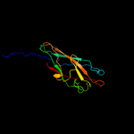
| 1 |
|
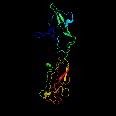
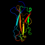
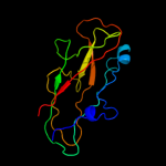
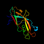
PDB 1klf chain P
Region: 118 - 411
Aligned: 208
Modelled: 234
Confidence: 99.8%
Identity: 21%
PDB header:chaperone/adhesin complex
Chain: P: PDB Molecule:fimh protein;
PDBTitle: fimh adhesin-fimc chaperone complex with d-mannose
Phyre2
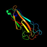
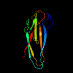
| 2 |
|
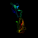
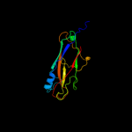
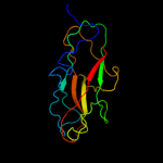
PDB 2jty chain A
Region: 245 - 411
Aligned: 142
Modelled: 148
Confidence: 99.8%
Identity: 25%
PDB header:structural protein
Chain: A: PDB Molecule:type-1 fimbrial protein, a chain;
PDBTitle: self-complemented variant of fima, the main subunit of type 1 pilus
Phyre2
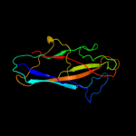
| 3 |
|
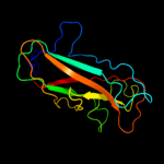
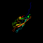
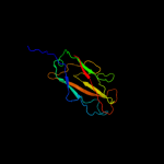
PDB 1pdk chain B
Region: 249 - 411
Aligned: 140
Modelled: 146
Confidence: 99.8%
Identity: 20%
Fold: Common fold of diphtheria toxin/transcription factors/cytochrome f
Superfamily: Bacterial adhesins
Family: Pilus subunits
Phyre2
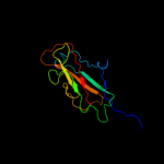
| 4 |
|
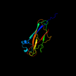
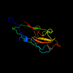
PDB 3jwn chain K
Region: 244 - 411
Aligned: 145
Modelled: 151
Confidence: 99.8%
Identity: 21%
PDB header:protein binding/cell adhesion
Chain: K: PDB Molecule:protein fimf;
PDBTitle: complex of fimc, fimf, fimg and fimh
Phyre2
| 5 |
|
PDB 3jwn chain L
Region: 244 - 411
Aligned: 145
Modelled: 151
Confidence: 99.8%
Identity: 21%
PDB header:protein binding/cell adhesion
Chain: L: PDB Molecule:protein fimf;
PDBTitle: complex of fimc, fimf, fimg and fimh
Phyre2
| 6 |
|
PDB 3jwn chain E
Region: 244 - 411
Aligned: 145
Modelled: 151
Confidence: 99.8%
Identity: 21%
PDB header:protein binding/cell adhesion
Chain: E: PDB Molecule:protein fimf;
PDBTitle: complex of fimc, fimf, fimg and fimh
Phyre2
| 7 |
|
PDB 3jwn chain F
Region: 244 - 411
Aligned: 145
Modelled: 151
Confidence: 99.8%
Identity: 21%
PDB header:protein binding/cell adhesion
Chain: F: PDB Molecule:protein fimf;
PDBTitle: complex of fimc, fimf, fimg and fimh
Phyre2
| 8 |
|
PDB 3bfw chain A
Region: 251 - 411
Aligned: 128
Modelled: 141
Confidence: 99.8%
Identity: 17%
PDB header:structural protein/structural protein
Chain: A: PDB Molecule:protein fimg;
PDBTitle: crystal structure of truncated fimg (fimgt) in complex with the donor2 strand peptide of fimf (dsf)
Phyre2
| 9 |
|
PDB 3bwu chain F
Region: 260 - 411
Aligned: 126
Modelled: 135
Confidence: 99.8%
Identity: 25%
PDB header:chaperone, structural, membrane protein
Chain: F: PDB Molecule:protein fimf;
PDBTitle: crystal structure of the ternary complex of fimd (n-terminal domain,2 fimdn) with fimc and the n-terminally truncated pilus subunit fimf3 (fimft)
Phyre2
| 10 |
|
PDB 2j2z chain B domain 1
Region: 244 - 411
Aligned: 142
Modelled: 147
Confidence: 99.8%
Identity: 18%
Fold: Common fold of diphtheria toxin/transcription factors/cytochrome f
Superfamily: Bacterial adhesins
Family: Pilus subunits
Phyre2
| 11 |
|
PDB 1ze3 chain H domain 1
Region: 252 - 411
Aligned: 120
Modelled: 138
Confidence: 99.7%
Identity: 18%
Fold: Common fold of diphtheria toxin/transcription factors/cytochrome f
Superfamily: Bacterial adhesins
Family: Pilus subunits
Phyre2
| 12 |
|
PDB 2jmr chain A
Region: 245 - 411
Aligned: 144
Modelled: 150
Confidence: 99.7%
Identity: 22%
PDB header:cell adhesion
Chain: A: PDB Molecule:fimf;
PDBTitle: nmr structure of the e. coli type 1 pilus subunit fimf
Phyre2
| 13 |
|
PDB 2uy6 chain B domain 1
Region: 244 - 411
Aligned: 143
Modelled: 143
Confidence: 99.7%
Identity: 21%
Fold: Common fold of diphtheria toxin/transcription factors/cytochrome f
Superfamily: Bacterial adhesins
Family: Pilus subunits
Phyre2
| 14 |
|
PDB 2w07 chain B
Region: 248 - 411
Aligned: 120
Modelled: 133
Confidence: 99.7%
Identity: 20%
PDB header:cell adhesion
Chain: B: PDB Molecule:minor pilin subunit papf;
PDBTitle: structural determinants of polymerization reactivity of the2 p pilus adaptor subunit papf
Phyre2
| 15 |
|
PDB 1n12 chain A
Region: 252 - 411
Aligned: 135
Modelled: 146
Confidence: 99.3%
Identity: 16%
Fold: Common fold of diphtheria toxin/transcription factors/cytochrome f
Superfamily: Bacterial adhesins
Family: Pilus subunits
Phyre2
| 16 |
|
PDB 2wmp chain B
Region: 268 - 412
Aligned: 112
Modelled: 116
Confidence: 51.6%
Identity: 17%
PDB header:chaperone
Chain: B: PDB Molecule:papg protein;
PDBTitle: structure of the e. coli chaperone papd in complex with the pilin2 domain of the papgii adhesin
Phyre2
| 17 |
|
PDB 3cl5 chain A
Region: 45 - 68
Aligned: 23
Modelled: 23
Confidence: 15.2%
Identity: 26%
PDB header:hydrolase
Chain: A: PDB Molecule:hemagglutinin-esterase;
PDBTitle: structure of coronavirus hemagglutinin-esterase in complex with 4,9-o-2 diacetyl sialic acid
Phyre2
| 18 |
|
PDB 3nqn chain B
Region: 378 - 400
Aligned: 23
Modelled: 23
Confidence: 7.6%
Identity: 13%
PDB header:structural genomics, unknown function
Chain: B: PDB Molecule:uncharacterized protein;
PDBTitle: crystal structure of a protein with unknown function. (dr_2006) from2 deinococcus radiodurans at 1.88 a resolution
Phyre2
| 19 |
|
PDB 3c7b chain B domain 3
Region: 253 - 298
Aligned: 43
Modelled: 46
Confidence: 7.3%
Identity: 21%
Fold: Nitrite and sulphite reductase 4Fe-4S domain-like
Superfamily: Nitrite and sulphite reductase 4Fe-4S domain-like
Family: Nitrite and sulphite reductase 4Fe-4S domain-like
Phyre2
| 20 |
|
PDB 2akj chain A domain 4
Region: 285 - 298
Aligned: 14
Modelled: 14
Confidence: 6.6%
Identity: 7%
Fold: Nitrite and sulphite reductase 4Fe-4S domain-like
Superfamily: Nitrite and sulphite reductase 4Fe-4S domain-like
Family: Nitrite and sulphite reductase 4Fe-4S domain-like
Phyre2
| 21 |
|
| 22 |
|
| 23 |
|
| 24 |
|