Phyre Home Retrieve Phyre Job Id
Emaill.a.kelley@imperial.ac.uk
DescriptionP52107
DateThu Jan 5 12:05:23 GMT 2012
Unique Job ID1dc6584a5132af1e

Summary 

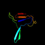
Top model
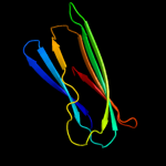
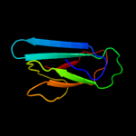
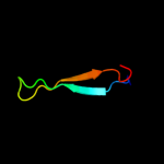
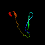
Image coloured by rainbow N → C terminus
Model (left) based on template c2xskA_
Top template information
PDB header:chaperone
Chain: A: PDB Molecule:csgc;
PDBTitle: e. coli curli protein csgc - secys
Confidence and coverage
Confidence:100.0% Coverage: 85%
93 residues ( 85% of your sequence) have been modelled with 100.0% confidence by the single highest scoring template.
3D viewing
Interactive 3D view in Jmol

Sequence analysis 

Secondary structure and disorder prediction 

   1........10.........20.........30.........40.........50.........60
Sequence  MNTLLLLAALSSQITFNTTQQGDVYTIIPEVTLTQSCLCRVQILSLREGSSGQSQTKQEK
Secondary structure 


SS confidence 


Disorder  ????

??
???


???????????????
Disorder confidence 


   .........70.........80.........90.........100.........110
Sequence  TLSLPANQPIALTKLSLNISPDDRVKIVVTVSDGQSLHLSQQWPPSSEKS
Secondary structure 


SS confidence 


Disorder 


?
???


?


?


???????????
Disorder confidence 


Confidence Key
High(9)                    Low (0)
?Disordered
Alpha helix
Beta strand

Domain analysis 

Hover over an aligned region to see model and summary info

Please note, only up to the top 20 hits are modelled to reduce computer load

RankAligned region

PDB 2xsk chain A

3D model

Region: 11 - 105
Aligned: 93
Modelled: 95
Confidence: 100.0%
Identity: 98%
PDB header:chaperone
Chain: A: PDB Molecule:csgc;
PDBTitle: e. coli curli protein csgc - secys

Phyre2

PDB 1x9l chain A

3D model

Region: 33 - 100
Aligned: 67
Modelled: 68
Confidence: 42.4%
Identity: 19%
Fold: Common fold of diphtheria toxin/transcription factors/cytochrome f
Superfamily: DR1885-like metal-binding protein
Family: DR1885-like metal-binding protein

Phyre2

PDB 1x99 chain A

3D model

Region: 11 - 100
Aligned: 88
Modelled: 89
Confidence: 42.1%
Identity: 16%
Fold: Cytolysin/lectin
Superfamily: Cytolysin/lectin
Family: Fungal fruit body lectin

Phyre2

PDB 2ofe chain A

3D model

Region: 11 - 100
Aligned: 88
Modelled: 90
Confidence: 33.1%
Identity: 15%
PDB header:sugar binding protein
Chain: A: PDB Molecule:sclerotium rolfsii lectin;
PDBTitle: the crystal structure of sclerotium rolfsii lectin in complex with n-2 acetyl-d-glucosamine

Phyre2

PDB 2k6z chain A

3D model

Region: 31 - 100
Aligned: 70
Modelled: 70
Confidence: 30.9%
Identity: 14%
PDB header:metal transport
Chain: A: PDB Molecule:putative uncharacterized protein ttha1943;
PDBTitle: solution structures of copper loaded form pcua (trans2 conformation of the peptide bond involving the nitrogen of3 p14)

Phyre2

PDB 2yhg chain A

3D model

Region: 56 - 100
Aligned: 33
Modelled: 45
Confidence: 16.2%
Identity: 21%
PDB header:hydrolase
Chain: A: PDB Molecule:cellulose-binding protein;
PDBTitle: ab initio phasing of a nucleoside hydrolase-related hypothetical2 protein from saccharophagus degradans that is associated with3 carbohydrate metabolism

Phyre2

PDB 1dki chain A

3D model

Region: 76 - 97
Aligned: 22
Modelled: 22
Confidence: 16.1%
Identity: 23%
Fold: Cysteine proteinases
Superfamily: Cysteine proteinases
Family: Papain-like

Phyre2

PDB 1ex0 chain A domain 1

3D model

Region: 47 - 86
Aligned: 40
Modelled: 40
Confidence: 15.5%
Identity: 13%
Fold: Immunoglobulin-like beta-sandwich
Superfamily: E set domains
Family: Transglutaminase N-terminal domain

Phyre2

PDB 2wda chain A

3D model

Region: 31 - 100
Aligned: 70
Modelled: 70
Confidence: 15.0%
Identity: 14%
PDB header:lyase
Chain: A: PDB Molecule:putative secreted lyase;
PDBTitle: the x-ray structure of the streptomyces coelicolor a32 chondroitin ac lyase in complex with chondroitin sulphate

Phyre2

PDB 2e7m chain A

3D model

Region: 49 - 103
Aligned: 49
Modelled: 55
Confidence: 13.7%
Identity: 33%
PDB header:structural protein
Chain: A: PDB Molecule:protein kiaa0319;
PDBTitle: solution structure of the pkd domain (329-428) from human2 kiaa0319

Phyre2

PDB 1pvj chain A

3D model

Region: 76 - 97
Aligned: 22
Modelled: 22
Confidence: 13.7%
Identity: 23%
Fold: Cysteine proteinases
Superfamily: Cysteine proteinases
Family: Papain-like

Phyre2

PDB 3bb7 chain A

3D model

Region: 76 - 97
Aligned: 22
Modelled: 22
Confidence: 12.3%
Identity: 27%
PDB header:hydrolase
Chain: A: PDB Molecule:interpain a;
PDBTitle: structure of prevotella intermedia prointerpain a fragment 39-3592 (mutant c154a)

Phyre2

PDB 2kj6 chain A

3D model

Region: 80 - 107
Aligned: 28
Modelled: 28
Confidence: 12.2%
Identity: 21%
PDB header:chaperone
Chain: A: PDB Molecule:tubulin folding cofactor b;
PDBTitle: nmr solution structure of a tubulin folding cofactor b2 obtained from arabidopsis thaliana: northeast structural3 genomics consortium target ar3436a

Phyre2

PDB 1w8o chain A domain 1

3D model

Region: 76 - 100
Aligned: 25
Modelled: 25
Confidence: 12.2%
Identity: 16%
Fold: Immunoglobulin-like beta-sandwich
Superfamily: E set domains
Family: E-set domains of sugar-utilizing enzymes

Phyre2

PDB 2ys4 chain A

3D model

Region: 49 - 108
Aligned: 59
Modelled: 60
Confidence: 11.1%
Identity: 22%
PDB header:structural genomics, unknown function
Chain: A: PDB Molecule:hydrocephalus-inducing protein homolog;
PDBTitle: solution structure of the n-terminal papd-like domain of2 hydin protein from human

Phyre2

PDB 1vjj chain A domain 1

3D model

Region: 38 - 86
Aligned: 49
Modelled: 49
Confidence: 10.4%
Identity: 16%
Fold: Immunoglobulin-like beta-sandwich
Superfamily: E set domains
Family: Transglutaminase N-terminal domain

Phyre2

PDB 2q3z chain A domain 1

3D model

Region: 48 - 86
Aligned: 39
Modelled: 39
Confidence: 10.3%
Identity: 36%
Fold: Immunoglobulin-like beta-sandwich
Superfamily: E set domains
Family: Transglutaminase N-terminal domain

Phyre2

PDB 1z8g chain A domain 2

3D model

Region: 40 - 54
Aligned: 15
Modelled: 15
Confidence: 10.0%
Identity: 20%
Fold: SRCR-like
Superfamily: SRCR-like
Family: Hepsin, N-terminal domain

Phyre2

PDB 2ezv chain A

3D model

Region: 46 - 78
Aligned: 33
Modelled: 33
Confidence: 8.3%
Identity: 27%
PDB header:hydrolase/dna
Chain: A: PDB Molecule:type ii restriction enzyme sfii;
PDBTitle: crystal structure of tetrameric restriction endonuclease2 sfii bound to cognate dna.

Phyre2

PDB 1ekr chain A

3D model

Region: 64 - 92
Aligned: 29
Modelled: 29
Confidence: 7.8%
Identity: 31%
Fold: Ferredoxin-like
Superfamily: Molybdenum cofactor biosynthesis protein C, MoaC
Family: Molybdenum cofactor biosynthesis protein C, MoaC

Phyre2
1

c2xskA_
2

d1x9la_
3

d1x99a_
4

c2ofeA_
5

c2k6zA_
6

c2yhgA_
7

d1dkia_
8

d1ex0a1
9

c2wdaA_
10

c2e7mA_
11

d1pvja_
12

c3bb7A_
13

c2kj6A_
14

d1w8oa1
15

c2ys4A_
16

d1vjja1
17

d2q3za1
18

d1z8ga2
19

c2ezvA_
20

d1ekra_
21


22


23


24


25


26


27


28


29


Detailed template information 

#
Template Alignment Coverage3D Model Confidence
% i.d. Template Information
1c2xskA_


100.0 98 PDB header:chaperone
Chain: A: PDB Molecule:csgc;
PDBTitle: e. coli curli protein csgc - secys
2d1x9la_


42.4 19 Fold:Common fold of diphtheria toxin/transcription factors/cytochrome f
Superfamily:DR1885-like metal-binding protein
Family:DR1885-like metal-binding protein
3d1x99a_


42.1 16 Fold:Cytolysin/lectin
Superfamily:Cytolysin/lectin
Family:Fungal fruit body lectin
4c2ofeA_


33.1 15 PDB header:sugar binding protein
Chain: A: PDB Molecule:sclerotium rolfsii lectin;
PDBTitle: the crystal structure of sclerotium rolfsii lectin in complex with n-2 acetyl-d-glucosamine
5c2k6zA_


30.9 14 PDB header:metal transport
Chain: A: PDB Molecule:putative uncharacterized protein ttha1943;
PDBTitle: solution structures of copper loaded form pcua (trans2 conformation of the peptide bond involving the nitrogen of3 p14)
6c2yhgA_


16.2 21 PDB header:hydrolase
Chain: A: PDB Molecule:cellulose-binding protein;
PDBTitle: ab initio phasing of a nucleoside hydrolase-related hypothetical2 protein from saccharophagus degradans that is associated with3 carbohydrate metabolism
7d1dkia_


16.1 23 Fold:Cysteine proteinases
Superfamily:Cysteine proteinases
Family:Papain-like
8d1ex0a1


15.5 13 Fold:Immunoglobulin-like beta-sandwich
Superfamily:E set domains
Family:Transglutaminase N-terminal domain
9c2wdaA_


15.0 14 PDB header:lyase
Chain: A: PDB Molecule:putative secreted lyase;
PDBTitle: the x-ray structure of the streptomyces coelicolor a32 chondroitin ac lyase in complex with chondroitin sulphate
10c2e7mA_


13.7 33 PDB header:structural protein
Chain: A: PDB Molecule:protein kiaa0319;
PDBTitle: solution structure of the pkd domain (329-428) from human2 kiaa0319
11d1pvja_


13.7 23 Fold:Cysteine proteinases
Superfamily:Cysteine proteinases
Family:Papain-like
12c3bb7A_


12.3 27 PDB header:hydrolase
Chain: A: PDB Molecule:interpain a;
PDBTitle: structure of prevotella intermedia prointerpain a fragment 39-3592 (mutant c154a)
13c2kj6A_


12.2 21 PDB header:chaperone
Chain: A: PDB Molecule:tubulin folding cofactor b;
PDBTitle: nmr solution structure of a tubulin folding cofactor b2 obtained from arabidopsis thaliana: northeast structural3 genomics consortium target ar3436a
14d1w8oa1


12.2 16 Fold:Immunoglobulin-like beta-sandwich
Superfamily:E set domains
Family:E-set domains of sugar-utilizing enzymes
15c2ys4A_


11.1 22 PDB header:structural genomics, unknown function
Chain: A: PDB Molecule:hydrocephalus-inducing protein homolog;
PDBTitle: solution structure of the n-terminal papd-like domain of2 hydin protein from human
16d1vjja1


10.4 16 Fold:Immunoglobulin-like beta-sandwich
Superfamily:E set domains
Family:Transglutaminase N-terminal domain
17d2q3za1


10.3 36 Fold:Immunoglobulin-like beta-sandwich
Superfamily:E set domains
Family:Transglutaminase N-terminal domain
18d1z8ga2


10.0 20 Fold:SRCR-like
Superfamily:SRCR-like
Family:Hepsin, N-terminal domain
19c2ezvA_


8.3 27 PDB header:hydrolase/dna
Chain: A: PDB Molecule:type ii restriction enzyme sfii;
PDBTitle: crystal structure of tetrameric restriction endonuclease2 sfii bound to cognate dna.
20d1ekra_


7.8 31 Fold:Ferredoxin-like
Superfamily:Molybdenum cofactor biosynthesis protein C, MoaC
Family:Molybdenum cofactor biosynthesis protein C, MoaC
21c3h8dC_


not modelled 7.8 29 PDB header:motor protein/signaling protein
Chain: C: PDB Molecule:myosin-vi;
PDBTitle: crystal structure of myosin vi in complex with dab2 peptide
22c2ideE_


not modelled 7.4 31 PDB header:biosynthetic protein
Chain: E: PDB Molecule:molybdenum cofactor biosynthesis protein c;
PDBTitle: crystal structure of the molybdenum cofactor biosynthesis protein c2 (ttha1789) from thermus theromophilus hb8
23c2xr4A_


not modelled 6.9 17 PDB header:sugar binding protein
Chain: A: PDB Molecule:lectin;
PDBTitle: c-terminal domain of bc2l-c lectin from burkholderia cenocepacia
24d2zkmx2


not modelled 6.7 23 Fold:C2 domain-like
Superfamily:C2 domain (Calcium/lipid-binding domain, CaLB)
Family:PLC-like (P variant)
25d1g0da1


not modelled 5.7 22 Fold:Immunoglobulin-like beta-sandwich
Superfamily:E set domains
Family:Transglutaminase N-terminal domain
26c2zq0B_


not modelled 5.6 19 PDB header:hydrolase
Chain: B: PDB Molecule:alpha-glucosidase (alpha-glucosidase susb);
PDBTitle: crystal structure of susb complexed with acarbose
27c2jnzA_


not modelled 5.2 9 PDB header:allergen
Chain: A: PDB Molecule:phl p 3 allergen;
PDBTitle: solution structure of phl p 3, a major allergen from2 timothy grass pollen
28d1v7wa2


not modelled 5.1 18 Fold:Supersandwich
Superfamily:Galactose mutarotase-like
Family:Glycosyltransferase family 36 N-terminal domain
29c2emcA_


not modelled 5.1 64 PDB header:transcription
Chain: A: PDB Molecule:zinc finger protein 473;
PDBTitle: solution structure of the c2h2 type zinc finger (region 641-2 673) of human zinc finger protein 473

Binding site prediction 

Due to computational demand, binding site predictions are not run for batch jobs

If you want to predict binding sites, please manually submit your model of choice to 3DLigandSite


Phyre is for academic use only

Please cite: Protein structure prediction on the web: a case study using the Phyre server
Kelley LA and Sternberg MJE. Nature Protocols 4, 363 - 371 (2009) [pdf] [Import into BibTeX]
 
If you use the binding site predictions from 3DLigandSite, please also cite:
3DLigandSite: predicting ligand-binding sites using similar structures.
Wass MN, Kelley LA and Sternberg MJ Nucleic Acids Research 38, W469-73 (2010) [PubMed]
 
© Structural Bioinformatics Group
Imperial College London
Lawrence Kelley, Benjamin Jefferys
Disclaimer
Terms and Conditions
Component software
Template detection: HHpred 1.51
Secondary structure prediction: Psi-pred 2.5
Disorder prediction: Disopred 2.4
Transmembrane prediction: Memsat_SVM
Multi-template modelling and ab initio: Poing 1.0