| 1 |
|
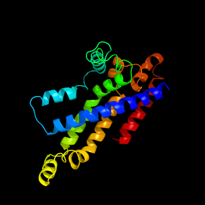
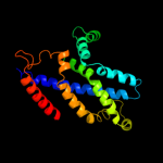
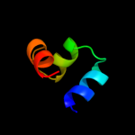
PDB 3d31 chain C domain 1
Region: 6 - 209
Aligned: 204
Modelled: 204
Confidence: 100.0%
Identity: 13%
Fold: MetI-like
Superfamily: MetI-like
Family: MetI-like
Phyre2
| 2 |
|
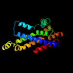
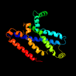
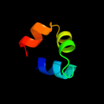
PDB 3d31 chain D
Region: 6 - 209
Aligned: 204
Modelled: 204
Confidence: 100.0%
Identity: 13%
PDB header:transport protein
Chain: D: PDB Molecule:sulfate/molybdate abc transporter, permease
PDBTitle: modbc from methanosarcina acetivorans
Phyre2
| 3 |
|
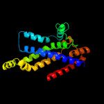
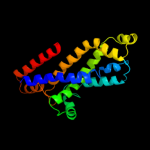
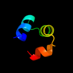
PDB 2onk chain C domain 1
Region: 6 - 211
Aligned: 204
Modelled: 206
Confidence: 100.0%
Identity: 13%
Fold: MetI-like
Superfamily: MetI-like
Family: MetI-like
Phyre2
| 4 |
|
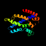
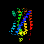
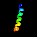
PDB 2onk chain C
Region: 6 - 211
Aligned: 204
Modelled: 206
Confidence: 100.0%
Identity: 13%
PDB header:membrane protein
Chain: C: PDB Molecule:molybdate/tungstate abc transporter, permease
PDBTitle: abc transporter modbc in complex with its binding protein2 moda
Phyre2
| 5 |
|
PDB 2r6g chain F
Region: 8 - 207
Aligned: 200
Modelled: 199
Confidence: 100.0%
Identity: 15%
PDB header:hydrolase/transport protein
Chain: F: PDB Molecule:maltose transport system permease protein malf;
PDBTitle: the crystal structure of the e. coli maltose transporter
Phyre2
| 6 |
|
PDB 2r6g chain F domain 2
Region: 3 - 207
Aligned: 205
Modelled: 205
Confidence: 100.0%
Identity: 12%
Fold: MetI-like
Superfamily: MetI-like
Family: MetI-like
Phyre2
| 7 |
|
PDB 3fh6 chain F
Region: 3 - 214
Aligned: 212
Modelled: 211
Confidence: 100.0%
Identity: 13%
PDB header:transport protein
Chain: F: PDB Molecule:maltose transport system permease protein malf;
PDBTitle: crystal structure of the resting state maltose transporter from e.2 coli
Phyre2
| 8 |
|
PDB 3dhw chain A domain 1
Region: 7 - 207
Aligned: 198
Modelled: 201
Confidence: 99.9%
Identity: 13%
Fold: MetI-like
Superfamily: MetI-like
Family: MetI-like
Phyre2
| 9 |
|
PDB 2r6g chain G domain 1
Region: 8 - 222
Aligned: 209
Modelled: 215
Confidence: 99.9%
Identity: 12%
Fold: MetI-like
Superfamily: MetI-like
Family: MetI-like
Phyre2
| 10 |
|
PDB 2jwa chain A
Region: 13 - 46
Aligned: 34
Modelled: 34
Confidence: 32.1%
Identity: 18%
PDB header:transferase
Chain: A: PDB Molecule:receptor tyrosine-protein kinase erbb-2;
PDBTitle: erbb2 transmembrane segment dimer spatial structure
Phyre2
| 11 |
|
PDB 1umq chain A
Region: 108 - 139
Aligned: 32
Modelled: 32
Confidence: 22.8%
Identity: 13%
PDB header:dna-binding protein
Chain: A: PDB Molecule:photosynthetic apparatus regulatory protein;
PDBTitle: solution structure and dna binding of the effector domain2 from the global regulator prra(rega) from r. sphaeroides:3 insights into dna binding specificity
Phyre2
| 12 |
|
PDB 1umq chain A
Region: 108 - 139
Aligned: 32
Modelled: 32
Confidence: 22.8%
Identity: 13%
Fold: DNA/RNA-binding 3-helical bundle
Superfamily: Homeodomain-like
Family: FIS-like
Phyre2
| 13 |
|
PDB 1ntc chain A
Region: 108 - 139
Aligned: 32
Modelled: 32
Confidence: 21.2%
Identity: 22%
Fold: DNA/RNA-binding 3-helical bundle
Superfamily: Homeodomain-like
Family: FIS-like
Phyre2
| 14 |
|
PDB 1fip chain A
Region: 108 - 139
Aligned: 32
Modelled: 32
Confidence: 19.3%
Identity: 13%
Fold: DNA/RNA-binding 3-helical bundle
Superfamily: Homeodomain-like
Family: FIS-like
Phyre2
| 15 |
|
PDB 3e7l chain D
Region: 108 - 139
Aligned: 32
Modelled: 32
Confidence: 17.4%
Identity: 6%
PDB header:transcription regulator
Chain: D: PDB Molecule:transcriptional regulator (ntrc family);
PDBTitle: crystal structure of sigma54 activator ntrc4's dna binding2 domain
Phyre2
| 16 |
|
PDB 1eto chain B
Region: 108 - 139
Aligned: 32
Modelled: 32
Confidence: 17.1%
Identity: 13%
Fold: DNA/RNA-binding 3-helical bundle
Superfamily: Homeodomain-like
Family: FIS-like
Phyre2
| 17 |
|
PDB 1etx chain A
Region: 108 - 139
Aligned: 32
Modelled: 32
Confidence: 16.5%
Identity: 9%
Fold: DNA/RNA-binding 3-helical bundle
Superfamily: Homeodomain-like
Family: FIS-like
Phyre2
| 18 |
|
PDB 2hx6 chain A
Region: 108 - 133
Aligned: 26
Modelled: 26
Confidence: 13.1%
Identity: 23%
PDB header:hydrolase
Chain: A: PDB Molecule:ribonuclease;
PDBTitle: solution structure analysis of the phage t42 endoribonuclease regb
Phyre2
| 19 |
|
PDB 2cw1 chain A
Region: 118 - 132
Aligned: 15
Modelled: 15
Confidence: 13.1%
Identity: 33%
PDB header:de novo protein
Chain: A: PDB Molecule:sn4m;
PDBTitle: solution structure of the de novo-designed lambda cro fold2 protein
Phyre2
| 20 |
|
PDB 1g2h chain A
Region: 108 - 139
Aligned: 31
Modelled: 32
Confidence: 10.7%
Identity: 19%
Fold: DNA/RNA-binding 3-helical bundle
Superfamily: Homeodomain-like
Family: FIS-like
Phyre2