Phyre Home Retrieve Phyre Job Id
Emaill.a.kelley@imperial.ac.uk
DescriptionP28247
DateThu Jan 5 11:44:32 GMT 2012
Unique Job ID19b77d83af5b0ff5

Summary 

Top model
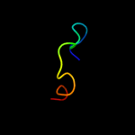
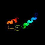
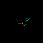
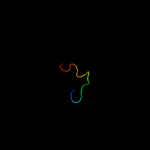
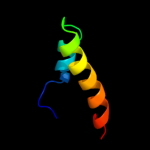
Image coloured by rainbow N → C terminus
Model (left) based on template c3jruB_
Top template information
PDB header:hydrolase
Chain: B: PDB Molecule:probable cytosol aminopeptidase;
PDBTitle: crystal structure of leucyl aminopeptidase (pepa) from xoo0834,2 xanthomonas oryzae pv. oryzae kacc10331
Confidence and coverage
Confidence: 30.5% Coverage: 10%
17 residues ( 10% of your sequence) have been modelled with 30.5% confidence by the single highest scoring template.
You may wish to submit your sequence to Phyrealarm. This will automatically scan your sequence every week for new potential templates as they appear in the Phyre2 library.
Please note: You must be registered and logged in to use Phyrealarm.
3D viewing
Interactive 3D view in Jmol

Sequence analysis 

Secondary structure and disorder prediction 

   1........10.........20.........30.........40.........50.........60
Sequence  MMARRSNAVRPLTIISHQHQTGGINIQPPRRMQFPRYRFIKKIEHRWVIRVVRGANIALR
Secondary structure 


SS confidence 


Disorder  ???????


??


????


Disorder confidence 


   .........70.........80.........90.........100.........110.........120
Sequence  LIEHEVTWTVLLRQRVAIVSDIMFRKQFERCITDDFAIDGDTIAADFTPGNSTANAELLC
Secondary structure 


SS confidence 


Disorder 


?


???
?


?


Disorder confidence 


   .........130.........140.........150.........160.......
Sequence  DKFIKSHVCDFACKNGGRALTRKSELLSAQYSGLITSLKGKRSMAYY
Secondary structure 


SS confidence 


Disorder 


????????


?

???????
Disorder confidence 


Confidence Key
High(9)                    Low (0)
?Disordered
Alpha helix
Beta strand

Domain analysis 

Hover over an aligned region to see model and summary info

Please note, only up to the top 20 hits are modelled to reduce computer load

RankAligned region

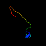
PDB 3jru chain B

3D model

Region: 21 - 37
Aligned: 17
Modelled: 17
Confidence: 30.5%
Identity: 29%
PDB header:hydrolase
Chain: B: PDB Molecule:probable cytosol aminopeptidase;
PDBTitle: crystal structure of leucyl aminopeptidase (pepa) from xoo0834,2 xanthomonas oryzae pv. oryzae kacc10331

Phyre2

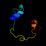
PDB 3ij3 chain A

3D model

Region: 5 - 37
Aligned: 33
Modelled: 33
Confidence: 29.6%
Identity: 18%
PDB header:hydrolase
Chain: A: PDB Molecule:cytosol aminopeptidase;
PDBTitle: 1.8 angstrom resolution crystal structure of cytosol aminopeptidase2 from coxiella burnetii

Phyre2

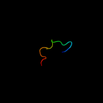
PDB 3h8g chain C

3D model

Region: 5 - 37
Aligned: 33
Modelled: 33
Confidence: 28.4%
Identity: 27%
PDB header:hydrolase
Chain: C: PDB Molecule:cytosol aminopeptidase;
PDBTitle: bestatin complex structure of leucine aminopeptidase from pseudomonas2 putida

Phyre2

PDB 1lan chain A

3D model

Region: 10 - 37
Aligned: 28
Modelled: 28
Confidence: 28.1%
Identity: 29%
PDB header:hydrolase (alpha-aminoacylpeptide)
Chain: A: PDB Molecule:leucine aminopeptidase;
PDBTitle: leucine aminopeptidase complex with l-leucinal

Phyre2

PDB 1v9v chain A domain 1

3D model

Region: 29 - 66
Aligned: 38
Modelled: 38
Confidence: 28.0%
Identity: 16%
Fold: Bromodomain-like
Superfamily: MAST3 pre-PK domain-like
Family: MAST3 pre-PK domain-like

Phyre2

PDB 1lam chain A domain 1

3D model

Region: 21 - 37
Aligned: 17
Modelled: 17
Confidence: 25.9%
Identity: 35%
Fold: Phosphorylase/hydrolase-like
Superfamily: Zn-dependent exopeptidases
Family: Leucine aminopeptidase, C-terminal domain

Phyre2

PDB 3bj4 chain B

3D model

Region: 59 - 86
Aligned: 25
Modelled: 28
Confidence: 22.1%
Identity: 36%
PDB header:signaling protein
Chain: B: PDB Molecule:potassium voltage-gated channel subfamily kqt
PDBTitle: the kcnq1 (kv7.1) c-terminus, a multi-tiered scaffold for2 subunit assembly and protein interaction

Phyre2

PDB 1gyt chain A domain 2

3D model

Region: 21 - 37
Aligned: 17
Modelled: 17
Confidence: 20.0%
Identity: 41%
Fold: Phosphorylase/hydrolase-like
Superfamily: Zn-dependent exopeptidases
Family: Leucine aminopeptidase, C-terminal domain

Phyre2

PDB 2hc9 chain A

3D model

Region: 21 - 37
Aligned: 17
Modelled: 17
Confidence: 18.1%
Identity: 29%
PDB header:hydrolase
Chain: A: PDB Molecule:leucine aminopeptidase 1;
PDBTitle: structure of caenorhabditis elegans leucine aminopeptidase-zinc2 complex (lap1)

Phyre2

PDB 1gyt chain G

3D model

Region: 21 - 37
Aligned: 17
Modelled: 17
Confidence: 17.7%
Identity: 41%
PDB header:hydrolase
Chain: G: PDB Molecule:cytosol aminopeptidase;
PDBTitle: e. coli aminopeptidase a (pepa)

Phyre2

PDB 3hfe chain C

3D model

Region: 59 - 81
Aligned: 20
Modelled: 23
Confidence: 16.9%
Identity: 45%
PDB header:transport protein
Chain: C: PDB Molecule:potassium voltage-gated channel subfamily kqt member 1;
PDBTitle: a trimeric form of the kv7.1 a domain tail

Phyre2

PDB 3h7h chain A

3D model

Region: 122 - 163
Aligned: 42
Modelled: 42
Confidence: 13.4%
Identity: 19%
PDB header:transcription
Chain: A: PDB Molecule:transcription elongation factor spt4;
PDBTitle: crystal structure of the human transcription elongation factor dsif,2 hspt4/hspt5 (176-273)

Phyre2

PDB 3kzw chain D

3D model

Region: 21 - 37
Aligned: 17
Modelled: 17
Confidence: 13.0%
Identity: 24%
PDB header:hydrolase
Chain: D: PDB Molecule:cytosol aminopeptidase;
PDBTitle: crystal structure of cytosol aminopeptidase from staphylococcus aureus2 col

Phyre2

PDB 3iwc chain D

3D model

Region: 3 - 24
Aligned: 21
Modelled: 22
Confidence: 12.8%
Identity: 29%
PDB header:lyase
Chain: D: PDB Molecule:s-adenosylmethionine decarboxylase;
PDBTitle: t. maritima adometdc complex with s-adenosylmethionine2 methyl ester

Phyre2

PDB 2obv chain A

3D model

Region: 75 - 137
Aligned: 62
Modelled: 63
Confidence: 12.6%
Identity: 16%
PDB header:transferase
Chain: A: PDB Molecule:s-adenosylmethionine synthetase isoform type-1;
PDBTitle: crystal structure of the human s-adenosylmethionine synthetase 1 in2 complex with the product

Phyre2

PDB 3kr5 chain E

3D model

Region: 21 - 37
Aligned: 17
Modelled: 17
Confidence: 12.5%
Identity: 24%
PDB header:hydrolase
Chain: E: PDB Molecule:m17 leucyl aminopeptidase;
PDBTitle: structure of a protease 4

Phyre2

PDB 2kzt chain A

3D model

Region: 116 - 153
Aligned: 38
Modelled: 38
Confidence: 12.1%
Identity: 29%
PDB header:apoptosis
Chain: A: PDB Molecule:programmed cell death protein 4;
PDBTitle: structure of the tandem ma-3 region of pdcd4

Phyre2

PDB 2d8x chain A domain 1

3D model

Region: 133 - 143
Aligned: 11
Modelled: 11
Confidence: 10.4%
Identity: 55%
Fold: Glucocorticoid receptor-like (DNA-binding domain)
Superfamily: Glucocorticoid receptor-like (DNA-binding domain)
Family: LIM domain

Phyre2

PDB 3iml chain B

3D model

Region: 71 - 137
Aligned: 66
Modelled: 67
Confidence: 9.9%
Identity: 20%
PDB header:transferase
Chain: B: PDB Molecule:s-adenosylmethionine synthetase;
PDBTitle: crystal structure of s-adenosylmethionine synthetase from burkholderia2 pseudomallei

Phyre2

PDB 1sr9 chain A domain 1

3D model

Region: 25 - 44
Aligned: 20
Modelled: 20
Confidence: 8.5%
Identity: 40%
Fold: RuvA C-terminal domain-like
Superfamily: post-HMGL domain-like
Family: DmpG/LeuA communication domain-like

Phyre2
1

c3jruB_
2

c3ij3A_
3

c3h8gC_
4

c1lanA_
5

d1v9va1
6

d1lama1
7

c3bj4B_
8

d1gyta2
9

c2hc9A_
10

c1gytG_
11

c3hfeC_
12

c3h7hA_
13

c3kzwD_
14

c3iwcD_
15

c2obvA_
16

c3kr5E_
17

c2kztA_
18

d2d8xa1
19

c3imlB_
20

d1sr9a1
21


22


23


24


25


26


27


28


29


30


31


32


33


34


35


36


37


38


39


40


Detailed template information 

#
Template Alignment Coverage3D Model Confidence
% i.d. Template Information
1c3jruB_


30.5 29 PDB header:hydrolase
Chain: B: PDB Molecule:probable cytosol aminopeptidase;
PDBTitle: crystal structure of leucyl aminopeptidase (pepa) from xoo0834,2 xanthomonas oryzae pv. oryzae kacc10331
2c3ij3A_


29.6 18 PDB header:hydrolase
Chain: A: PDB Molecule:cytosol aminopeptidase;
PDBTitle: 1.8 angstrom resolution crystal structure of cytosol aminopeptidase2 from coxiella burnetii
3c3h8gC_


28.4 27 PDB header:hydrolase
Chain: C: PDB Molecule:cytosol aminopeptidase;
PDBTitle: bestatin complex structure of leucine aminopeptidase from pseudomonas2 putida
4c1lanA_


28.1 29 PDB header:hydrolase (alpha-aminoacylpeptide)
Chain: A: PDB Molecule:leucine aminopeptidase;
PDBTitle: leucine aminopeptidase complex with l-leucinal
5d1v9va1


28.0 16 Fold:Bromodomain-like
Superfamily:MAST3 pre-PK domain-like
Family:MAST3 pre-PK domain-like
6d1lama1


25.9 35 Fold:Phosphorylase/hydrolase-like
Superfamily:Zn-dependent exopeptidases
Family:Leucine aminopeptidase, C-terminal domain
7c3bj4B_


22.1 36 PDB header:signaling protein
Chain: B: PDB Molecule:potassium voltage-gated channel subfamily kqt
PDBTitle: the kcnq1 (kv7.1) c-terminus, a multi-tiered scaffold for2 subunit assembly and protein interaction
8d1gyta2


20.0 41 Fold:Phosphorylase/hydrolase-like
Superfamily:Zn-dependent exopeptidases
Family:Leucine aminopeptidase, C-terminal domain
9c2hc9A_


18.1 29 PDB header:hydrolase
Chain: A: PDB Molecule:leucine aminopeptidase 1;
PDBTitle: structure of caenorhabditis elegans leucine aminopeptidase-zinc2 complex (lap1)
10c1gytG_


17.7 41 PDB header:hydrolase
Chain: G: PDB Molecule:cytosol aminopeptidase;
PDBTitle: e. coli aminopeptidase a (pepa)
11c3hfeC_


16.9 45 PDB header:transport protein
Chain: C: PDB Molecule:potassium voltage-gated channel subfamily kqt member 1;
PDBTitle: a trimeric form of the kv7.1 a domain tail
12c3h7hA_


13.4 19 PDB header:transcription
Chain: A: PDB Molecule:transcription elongation factor spt4;
PDBTitle: crystal structure of the human transcription elongation factor dsif,2 hspt4/hspt5 (176-273)
13c3kzwD_


13.0 24 PDB header:hydrolase
Chain: D: PDB Molecule:cytosol aminopeptidase;
PDBTitle: crystal structure of cytosol aminopeptidase from staphylococcus aureus2 col
14c3iwcD_


12.8 29 PDB header:lyase
Chain: D: PDB Molecule:s-adenosylmethionine decarboxylase;
PDBTitle: t. maritima adometdc complex with s-adenosylmethionine2 methyl ester
15c2obvA_


12.6 16 PDB header:transferase
Chain: A: PDB Molecule:s-adenosylmethionine synthetase isoform type-1;
PDBTitle: crystal structure of the human s-adenosylmethionine synthetase 1 in2 complex with the product
16c3kr5E_


12.5 24 PDB header:hydrolase
Chain: E: PDB Molecule:m17 leucyl aminopeptidase;
PDBTitle: structure of a protease 4
17c2kztA_


12.1 29 PDB header:apoptosis
Chain: A: PDB Molecule:programmed cell death protein 4;
PDBTitle: structure of the tandem ma-3 region of pdcd4
18d2d8xa1


10.4 55 Fold:Glucocorticoid receptor-like (DNA-binding domain)
Superfamily:Glucocorticoid receptor-like (DNA-binding domain)
Family:LIM domain
19c3imlB_


9.9 20 PDB header:transferase
Chain: B: PDB Molecule:s-adenosylmethionine synthetase;
PDBTitle: crystal structure of s-adenosylmethionine synthetase from burkholderia2 pseudomallei
20d1sr9a1


8.5 40 Fold:RuvA C-terminal domain-like
Superfamily:post-HMGL domain-like
Family:DmpG/LeuA communication domain-like
21c1mtpB_


not modelled 8.5 33 PDB header:structural genomics
Chain: B: PDB Molecule:serine proteinase inhibitor (serpin), chain b;
PDBTitle: the x-ray crystal structure of a serpin from a thermophilic2 prokaryote
22d2q49a2


not modelled 8.2 11 Fold:FwdE/GAPDH domain-like
Superfamily:Glyceraldehyde-3-phosphate dehydrogenase-like, C-terminal domain
Family:GAPDH-like
23d1edla_


not modelled 8.1 44 Fold:immunoglobulin/albumin-binding domain-like
Superfamily:Bacterial immunoglobulin/albumin-binding domains
Family:Immunoglobulin-binding protein A modules
24c2wshC_


not modelled 7.8 23 PDB header:hydrolase
Chain: C: PDB Molecule:endonuclease ii;
PDBTitle: structure of bacteriophage t4 endoii e118a mutant
25d1uc2a_


not modelled 7.8 16 Fold:Hypothetical protein PH1602
Superfamily:Hypothetical protein PH1602
Family:Hypothetical protein PH1602
26c2eloA_


not modelled 7.2 71 PDB header:transcription
Chain: A: PDB Molecule:zinc finger protein 406;
PDBTitle: solution structure of the 12th c2h2 zinc finger of human2 zinc finger protein 406
27d1vjpa1


not modelled 7.0 9 Fold:NAD(P)-binding Rossmann-fold domains
Superfamily:NAD(P)-binding Rossmann-fold domains
Family:Glyceraldehyde-3-phosphate dehydrogenase-like, N-terminal domain
28d1fc2c_


not modelled 6.7 41 Fold:immunoglobulin/albumin-binding domain-like
Superfamily:Bacterial immunoglobulin/albumin-binding domains
Family:Immunoglobulin-binding protein A modules
29d1ryqa_


not modelled 6.7 28 Fold:Rubredoxin-like
Superfamily:RNA polymerase subunits
Family:RpoE2-like
30d2db7a1


not modelled 6.5 18 Fold:Orange domain-like
Superfamily:Orange domain-like
Family:Hairy Orange domain
31c2zbtB_


not modelled 6.4 33 PDB header:lyase
Chain: B: PDB Molecule:pyridoxal biosynthesis lyase pdxs;
PDBTitle: crystal structure of pyridoxine biosynthesis protein from thermus2 thermophilus hb8
32c3so4C_


not modelled 6.3 16 PDB header:transferase
Chain: C: PDB Molecule:methionine-adenosyltransferase;
PDBTitle: methionine-adenosyltransferase from entamoeba histolytica
33c3femB_


not modelled 6.3 40 PDB header:biosynthetic protein, transferase
Chain: B: PDB Molecule:pyridoxine biosynthesis protein snz1;
PDBTitle: structure of the synthase subunit pdx1.1 (snz1) of plp synthase from2 saccharomyces cerevisiae
34d2p02a2


not modelled 6.1 47 Fold:S-adenosylmethionine synthetase
Superfamily:S-adenosylmethionine synthetase
Family:S-adenosylmethionine synthetase
35d1prtc2


not modelled 6.1 78 Fold:C-type lectin-like
Superfamily:C-type lectin-like
Family:Aerolysin/Pertussis toxin (APT) domain
36c2epgB_


not modelled 6.1 18 PDB header:structural genomics, unknown function
Chain: B: PDB Molecule:hypothetical protein ttha1785;
PDBTitle: crystal structure of ttha1785
37c2nv2U_


not modelled 5.9 33 PDB header:lyase/transferase
Chain: U: PDB Molecule:pyridoxal biosynthesis lyase pdxs;
PDBTitle: structure of the plp synthase complex pdx1/2 (yaad/e) from bacillus2 subtilis
38c3mtvA_


not modelled 5.8 25 PDB header:hydrolase
Chain: A: PDB Molecule:papain-like cysteine protease;
PDBTitle: the crystal structure of the prrsv nonstructural protein nsp1
39d1vkna2


not modelled 5.8 24 Fold:FwdE/GAPDH domain-like
Superfamily:Glyceraldehyde-3-phosphate dehydrogenase-like, C-terminal domain
Family:GAPDH-like
40c3rv2B_


not modelled 5.8 16 PDB header:transferase
Chain: B: PDB Molecule:s-adenosylmethionine synthase;
PDBTitle: crystal structure of s-adenosylmethionine synthetase from2 mycobacterium marinum

Binding site prediction 

Due to computational demand, binding site predictions are not run for batch jobs

If you want to predict binding sites, please manually submit your model of choice to 3DLigandSite


Phyre is for academic use only

Please cite: Protein structure prediction on the web: a case study using the Phyre server
Kelley LA and Sternberg MJE. Nature Protocols 4, 363 - 371 (2009) [pdf] [Import into BibTeX]
 
If you use the binding site predictions from 3DLigandSite, please also cite:
3DLigandSite: predicting ligand-binding sites using similar structures.
Wass MN, Kelley LA and Sternberg MJ Nucleic Acids Research 38, W469-73 (2010) [PubMed]
 
© Structural Bioinformatics Group
Imperial College London
Lawrence Kelley, Benjamin Jefferys
Disclaimer
Terms and Conditions
Component software
Template detection: HHpred 1.51
Secondary structure prediction: Psi-pred 2.5
Disorder prediction: Disopred 2.4
Transmembrane prediction: Memsat_SVM
Multi-template modelling and ab initio: Poing 1.0