Phyre Home Retrieve Phyre Job Id
Emaill.a.kelley@imperial.ac.uk
DescriptionP76418
DateThu Jan 5 12:22:53 GMT 2012
Unique Job ID17378e692b6701cd

Summary 

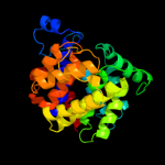
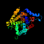
Top model
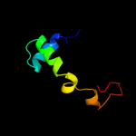
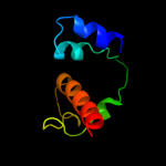
Image coloured by rainbow N → C terminus
Model (left) based on template c3g9dB_
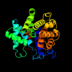
Top template information
PDB header:hydrolase
Chain: B: PDB Molecule:dinitrogenase reductase activacting
PDBTitle: crystal structure glycohydrolase
Confidence and coverage
Confidence:100.0% Coverage: 84%
282 residues ( 84% of your sequence) have been modelled with 100.0% confidence by the single highest scoring template.
Additional confident templates have been detected (see Domain analysis) which cover other regions of your sequence.
333 residues (100%) could be modelled at >90% confidence using multiple-templates.
You may wish to try resubmitting your sequence in "intensive" mode to model more of your sequence.
3D viewing
Interactive 3D view in Jmol

Sequence analysis 

Secondary structure and disorder prediction 

   1........10.........20.........30.........40.........50.........60
Sequence  MKTERILGALYGQALGDAMGMPSELWPRSRVKAHFGWIDRFLPGPKENNAACYFNRAEFT
Secondary structure 


SS confidence 


Disorder  ??


Disorder confidence 


   .........70.........80.........90.........100.........110.........120
Sequence  DDTSMALCLADALLEREGKIDPDLIGRNILDWALRFDAFNKNVLGPTSKIALNAIRDGKP
Secondary structure 


SS confidence 


Disorder 


Disorder confidence 


   .........130.........140.........150.........160.........170.........180
Sequence  VAELENNGVTNGAAMRVSPLGCLLPARDVDSFIDDVALASSPTHKSDLAVAGAVVIAWAI
Secondary structure 


SS confidence 


Disorder  ?????????


Disorder confidence 


   .........190.........200.........210.........220.........230.........240
Sequence  SRAIDGESWSAIVDSLPSIARHAQQKRITTFSASLAARLEIALKIVRNADGTESASEQLY
Secondary structure 


SS confidence 


Disorder 


Disorder confidence 


   .........250.........260.........270.........280.........290.........300
Sequence  QVVGAGTSTIESVPCAIALVELAQTDPNRCAVLCANLGGDTDTIGAMATAICGALHGVNA
Secondary structure 


SS confidence 


Disorder 


Disorder confidence 


   .........310.........320.........330....
Sequence  IDPALKAELDAVNQLDFNRYATALAKYRQQREAV
Secondary structure 


SS confidence 


Disorder 


????
Disorder confidence 


Confidence Key
High(9)                    Low (0)
?Disordered
Alpha helix
Beta strand

Domain analysis 

Hover over an aligned region to see model and summary info

Please note, only up to the top 20 hits are modelled to reduce computer load

RankAligned region

PDB 3g9d chain B

3D model

Region: 3 - 326
Aligned: 282
Modelled: 289
Confidence: 100.0%
Identity: 23%
PDB header:hydrolase
Chain: B: PDB Molecule:dinitrogenase reductase activacting
PDBTitle: crystal structure glycohydrolase

Phyre2

PDB 2woc chain A

3D model

Region: 3 - 328
Aligned: 284
Modelled: 284
Confidence: 100.0%
Identity: 23%
PDB header:hydrolase
Chain: A: PDB Molecule:adp-ribosyl-[dinitrogen reductase] glycohydrolase;
PDBTitle: crystal structure of the dinitrogenase reductase-activating2 glycohydrolase (drag) from rhodospirillum rubrum

Phyre2

PDB 1t5j chain A

3D model

Region: 3 - 330
Aligned: 297
Modelled: 312
Confidence: 100.0%
Identity: 29%
Fold: ADP-ribosylglycohydrolase
Superfamily: ADP-ribosylglycohydrolase
Family: ADP-ribosylglycohydrolase

Phyre2

PDB 3hfw chain A

3D model

Region: 4 - 334
Aligned: 321
Modelled: 331
Confidence: 100.0%
Identity: 20%
PDB header:hydrolase
Chain: A: PDB Molecule:protein adp-ribosylarginine hydrolase;
PDBTitle: crystal structure of human adp-ribosylhydrolase 1 (harh1)

Phyre2

PDB 2qty chain B

3D model

Region: 4 - 330
Aligned: 308
Modelled: 325
Confidence: 100.0%
Identity: 25%
PDB header:hydrolase
Chain: B: PDB Molecule:poly(adp-ribose) glycohydrolase arh3;
PDBTitle: crystal structure of mouse adp-ribosylhydrolase 3 (marh3)

Phyre2

PDB 2yzw chain A

3D model

Region: 2 - 328
Aligned: 279
Modelled: 304
Confidence: 100.0%
Identity: 26%
PDB header:hydrolase
Chain: A: PDB Molecule:adp-ribosylglycohydrolase;
PDBTitle: adp-ribosylglycohydrolase-related protein complex

Phyre2

PDB 2i7n chain A domain 2

3D model

Region: 246 - 304
Aligned: 57
Modelled: 59
Confidence: 19.1%
Identity: 14%
Fold: Ribonuclease H-like motif
Superfamily: Actin-like ATPase domain
Family: Fumble-like

Phyre2

PDB 1j9j chain A

3D model

Region: 276 - 330
Aligned: 51
Modelled: 55
Confidence: 16.8%
Identity: 24%
Fold: SurE-like
Superfamily: SurE-like
Family: SurE-like

Phyre2

PDB 2odb chain B

3D model

Region: 297 - 310
Aligned: 14
Modelled: 14
Confidence: 15.8%
Identity: 29%
PDB header:protein binding
Chain: B: PDB Molecule:serine/threonine-protein kinase pak 6;
PDBTitle: the crystal structure of human cdc42 in complex with the crib domain2 of human p21-activated kinase 6 (pak6)

Phyre2

PDB 1mw7 chain A

3D model

Region: 267 - 308
Aligned: 40
Modelled: 42
Confidence: 14.3%
Identity: 25%
Fold: YebC-like
Superfamily: YebC-like
Family: YebC-like

Phyre2

PDB 1e0a chain B

3D model

Region: 297 - 314
Aligned: 18
Modelled: 18
Confidence: 13.1%
Identity: 17%
PDB header:signalling protein
Chain: B: PDB Molecule:serine/threonine-protein kinase pak-alpha;
PDBTitle: cdc42 complexed with the gtpase binding domain of p212 activated kinase

Phyre2

PDB 3smp chain A

3D model

Region: 246 - 304
Aligned: 57
Modelled: 59
Confidence: 11.8%
Identity: 14%
PDB header:transferase
Chain: A: PDB Molecule:pantothenate kinase 1;
PDBTitle: monoclinic crystal structure of human pantothenate kinase 1 alpha

Phyre2

PDB 2i7p chain A domain 1

3D model

Region: 246 - 304
Aligned: 57
Modelled: 59
Confidence: 11.4%
Identity: 12%
Fold: Ribonuclease H-like motif
Superfamily: Actin-like ATPase domain
Family: Fumble-like

Phyre2

PDB 1f3m chain B

3D model

Region: 297 - 314
Aligned: 18
Modelled: 18
Confidence: 10.1%
Identity: 17%
PDB header:transferase
Chain: B: PDB Molecule:serine/threonine-protein kinase pak-alpha;
PDBTitle: crystal structure of human serine/threonine kinase pak1

Phyre2

PDB 1cee chain B

3D model

Region: 297 - 316
Aligned: 20
Modelled: 20
Confidence: 9.5%
Identity: 30%
PDB header:structural protein regulation
Chain: B: PDB Molecule:wiskott-aldrich syndrome protein wasp;
PDBTitle: solution structure of cdc42 in complex with the gtpase2 binding domain of wasp

Phyre2

PDB 2phj chain A

3D model

Region: 276 - 301
Aligned: 26
Modelled: 26
Confidence: 9.2%
Identity: 35%
PDB header:hydrolase
Chain: A: PDB Molecule:5'-nucleotidase sure;
PDBTitle: crystal structure of sure protein from aquifex aeolicus

Phyre2

PDB 2i7p chain A

3D model

Region: 246 - 304
Aligned: 57
Modelled: 59
Confidence: 8.6%
Identity: 12%
PDB header:transferase
Chain: A: PDB Molecule:pantothenate kinase 3;
PDBTitle: crystal structure of human pank3 in complex with accoa

Phyre2

PDB 2j0w chain A

3D model

Region: 279 - 294
Aligned: 16
Modelled: 16
Confidence: 8.3%
Identity: 25%
PDB header:transferase
Chain: A: PDB Molecule:lysine-sensitive aspartokinase 3;
PDBTitle: crystal structure of e. coli aspartokinase iii in complex2 with aspartate and adp (r-state)

Phyre2

PDB 2e6g chain I

3D model

Region: 276 - 301
Aligned: 26
Modelled: 26
Confidence: 8.3%
Identity: 38%
PDB header:hydrolase
Chain: I: PDB Molecule:5'-nucleotidase sure;
PDBTitle: crystal structure of the stationary phase survival protein sure from2 thermus thermophilus hb8 in complex with phosphate

Phyre2

PDB 3c9p chain A

3D model

Region: 254 - 326
Aligned: 63
Modelled: 68
Confidence: 8.2%
Identity: 17%
PDB header:structural genomics, unknown function
Chain: A: PDB Molecule:uncharacterized protein sp1917;
PDBTitle: crystal structure of uncharacterized protein sp1917

Phyre2
1

c3g9dB_
2

c2wocA_
3

d1t5ja_
4

c3hfwA_
5

c2qtyB_
6

c2yzwA_
7

d2i7na2
8

d1j9ja_
9

c2odbB_
10

d1mw7a_
11

c1e0aB_
12

c3smpA_
13

d2i7pa1
14

c1f3mB_
15

c1ceeB_
16

c2phjA_
17

c2i7pA_
18

c2j0wA_
19

c2e6gI_
20

c3c9pA_
21


22


23


24


25


26


27


28


29


30


31


32


33


34


35


36


37


38


Detailed template information 

#
Template Alignment Coverage3D Model Confidence
% i.d. Template Information
1c3g9dB_


100.0 23 PDB header:hydrolase
Chain: B: PDB Molecule:dinitrogenase reductase activacting
PDBTitle: crystal structure glycohydrolase
2c2wocA_


100.0 23 PDB header:hydrolase
Chain: A: PDB Molecule:adp-ribosyl-[dinitrogen reductase] glycohydrolase;
PDBTitle: crystal structure of the dinitrogenase reductase-activating2 glycohydrolase (drag) from rhodospirillum rubrum
3d1t5ja_


100.0 29 Fold:ADP-ribosylglycohydrolase
Superfamily:ADP-ribosylglycohydrolase
Family:ADP-ribosylglycohydrolase
4c3hfwA_


100.0 20 PDB header:hydrolase
Chain: A: PDB Molecule:protein adp-ribosylarginine hydrolase;
PDBTitle: crystal structure of human adp-ribosylhydrolase 1 (harh1)
5c2qtyB_


100.0 25 PDB header:hydrolase
Chain: B: PDB Molecule:poly(adp-ribose) glycohydrolase arh3;
PDBTitle: crystal structure of mouse adp-ribosylhydrolase 3 (marh3)
6c2yzwA_


100.0 26 PDB header:hydrolase
Chain: A: PDB Molecule:adp-ribosylglycohydrolase;
PDBTitle: adp-ribosylglycohydrolase-related protein complex
7d2i7na2


19.1 14 Fold:Ribonuclease H-like motif
Superfamily:Actin-like ATPase domain
Family:Fumble-like
8d1j9ja_


16.8 24 Fold:SurE-like
Superfamily:SurE-like
Family:SurE-like
9c2odbB_


15.8 29 PDB header:protein binding
Chain: B: PDB Molecule:serine/threonine-protein kinase pak 6;
PDBTitle: the crystal structure of human cdc42 in complex with the crib domain2 of human p21-activated kinase 6 (pak6)
10d1mw7a_


14.3 25 Fold:YebC-like
Superfamily:YebC-like
Family:YebC-like
11c1e0aB_


13.1 17 PDB header:signalling protein
Chain: B: PDB Molecule:serine/threonine-protein kinase pak-alpha;
PDBTitle: cdc42 complexed with the gtpase binding domain of p212 activated kinase
12c3smpA_


11.8 14 PDB header:transferase
Chain: A: PDB Molecule:pantothenate kinase 1;
PDBTitle: monoclinic crystal structure of human pantothenate kinase 1 alpha
13d2i7pa1


11.4 12 Fold:Ribonuclease H-like motif
Superfamily:Actin-like ATPase domain
Family:Fumble-like
14c1f3mB_


10.1 17 PDB header:transferase
Chain: B: PDB Molecule:serine/threonine-protein kinase pak-alpha;
PDBTitle: crystal structure of human serine/threonine kinase pak1
15c1ceeB_


9.5 30 PDB header:structural protein regulation
Chain: B: PDB Molecule:wiskott-aldrich syndrome protein wasp;
PDBTitle: solution structure of cdc42 in complex with the gtpase2 binding domain of wasp
16c2phjA_


9.2 35 PDB header:hydrolase
Chain: A: PDB Molecule:5'-nucleotidase sure;
PDBTitle: crystal structure of sure protein from aquifex aeolicus
17c2i7pA_


8.6 12 PDB header:transferase
Chain: A: PDB Molecule:pantothenate kinase 3;
PDBTitle: crystal structure of human pank3 in complex with accoa
18c2j0wA_


8.3 25 PDB header:transferase
Chain: A: PDB Molecule:lysine-sensitive aspartokinase 3;
PDBTitle: crystal structure of e. coli aspartokinase iii in complex2 with aspartate and adp (r-state)
19c2e6gI_


8.3 38 PDB header:hydrolase
Chain: I: PDB Molecule:5'-nucleotidase sure;
PDBTitle: crystal structure of the stationary phase survival protein sure from2 thermus thermophilus hb8 in complex with phosphate
20c3c9pA_


8.2 17 PDB header:structural genomics, unknown function
Chain: A: PDB Molecule:uncharacterized protein sp1917;
PDBTitle: crystal structure of uncharacterized protein sp1917
21c1ir6A_


not modelled 8.0 13 PDB header:hydrolase
Chain: A: PDB Molecule:exonuclease recj;
PDBTitle: crystal structure of exonuclease recj bound to manganese
22d1ir6a_


not modelled 8.0 13 Fold:DHH phosphoesterases
Superfamily:DHH phosphoesterases
Family:Exonuclease RecJ
23c3bowC_


not modelled 7.8 25 PDB header:hydrolase/hydrolase inhibitor
Chain: C: PDB Molecule:calpastatin;
PDBTitle: structure of m-calpain in complex with calpastatin
24d2j0wa1


not modelled 7.7 25 Fold:Carbamate kinase-like
Superfamily:Carbamate kinase-like
Family:PyrH-like
25c3devB_


not modelled 7.5 9 PDB header:structural genomics, unknown function
Chain: B: PDB Molecule:sh1221;
PDBTitle: crystal structure of sh1221 protein from staphylococcus haemolyticus,2 northeast structural genomics consortium target shr87
26c1w6kA_


not modelled 6.8 13 PDB header:isomerase
Chain: A: PDB Molecule:lanosterol synthase;
PDBTitle: structure of human osc in complex with lanosterol
27c2ogxB_


not modelled 6.7 18 PDB header:metal binding protein
Chain: B: PDB Molecule:molybdenum storage protein subunit beta;
PDBTitle: the crystal structure of the molybdenum storage protein from2 azotobacter vinelandii loaded with polyoxotungstates (wsto)
28d1ykwa1


not modelled 6.6 21 Fold:TIM beta/alpha-barrel
Superfamily:RuBisCo, C-terminal domain
Family:RuBisCo, large subunit, C-terminal domain
29c2k42A_


not modelled 5.8 14 PDB header:signaling protein
Chain: A: PDB Molecule:wiskott-aldrich syndrome protein;
PDBTitle: solution structure of the gtpase binding domain of wasp in2 complex with espfu, an ehec effector
30c3ab4K_


not modelled 5.7 31 PDB header:transferase
Chain: K: PDB Molecule:aspartokinase;
PDBTitle: crystal structure of feedback inhibition resistant mutant of aspartate2 kinase from corynebacterium glutamicum in complex with lysine and3 threonine
31c2ap7A_


not modelled 5.6 43 PDB header:antibiotic
Chain: A: PDB Molecule:bombinin h2;
PDBTitle: solution structure of bombinin h2 in dpc micelles
32d2cdqa1


not modelled 5.6 19 Fold:Carbamate kinase-like
Superfamily:Carbamate kinase-like
Family:PyrH-like
33c2v4oB_


not modelled 5.6 38 PDB header:hydrolase
Chain: B: PDB Molecule:multifunctional protein sur e;
PDBTitle: crystal structure of salmonella typhimurium sure at 2.752 angstrom resolution in monoclinic form
34d1w6ka1


not modelled 5.5 10 Fold:alpha/alpha toroid
Superfamily:Terpenoid cyclases/Protein prenyltransferases
Family:Terpene synthases
35d2gpia1


not modelled 5.4 10 Fold:Shew3726-like
Superfamily:Shew3726-like
Family:Shew3726-like
36d2hmfa1


not modelled 5.3 25 Fold:Carbamate kinase-like
Superfamily:Carbamate kinase-like
Family:PyrH-like
37d1ekqa_


not modelled 5.3 19 Fold:Ribokinase-like
Superfamily:Ribokinase-like
Family:Thiamin biosynthesis kinases
38c3ll9A_


not modelled 5.0 6 PDB header:transferase
Chain: A: PDB Molecule:isopentenyl phosphate kinase;
PDBTitle: x-ray structures of isopentenyl phosphate kinase

Binding site prediction 

Due to computational demand, binding site predictions are not run for batch jobs

If you want to predict binding sites, please manually submit your model of choice to 3DLigandSite


Phyre is for academic use only

Please cite: Protein structure prediction on the web: a case study using the Phyre server
Kelley LA and Sternberg MJE. Nature Protocols 4, 363 - 371 (2009) [pdf] [Import into BibTeX]
 
If you use the binding site predictions from 3DLigandSite, please also cite:
3DLigandSite: predicting ligand-binding sites using similar structures.
Wass MN, Kelley LA and Sternberg MJ Nucleic Acids Research 38, W469-73 (2010) [PubMed]
 
© Structural Bioinformatics Group
Imperial College London
Lawrence Kelley, Benjamin Jefferys
Disclaimer
Terms and Conditions
Component software
Template detection: HHpred 1.51
Secondary structure prediction: Psi-pred 2.5
Disorder prediction: Disopred 2.4
Transmembrane prediction: Memsat_SVM
Multi-template modelling and ab initio: Poing 1.0