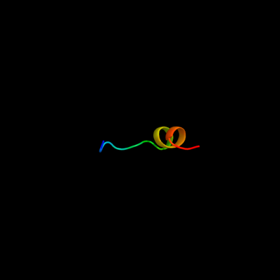
Phyre Home Retrieve Phyre Job Id
Emaill.a.kelley@imperial.ac.uk
DescriptionP52132
DateThu Jan 5 12:05:37 GMT 2012
Unique Job ID12f33cbd866485f6

Summary 

Top model
Image coloured by rainbow N → C terminus
Model (left) based on template c2f9jP_
Top template information
PDB header:rna binding protein
Chain: P: PDB Molecule:splicing factor 3b subunit 1;
PDBTitle: 3.0 angstrom resolution structure of a y22m mutant of the spliceosomal2 protein p14 bound to a region of sf3b155
Confidence and coverage
Confidence: 32.7% Coverage: 6%
16 residues ( 6% of your sequence) have been modelled with 32.7% confidence by the single highest scoring template.
You may wish to submit your sequence to Phyrealarm. This will automatically scan your sequence every week for new potential templates as they appear in the Phyre2 library.
Please note: You must be registered and logged in to use Phyrealarm.
3D viewing
Interactive 3D view in Jmol

Sequence analysis 

Secondary structure and disorder prediction 

   1........10.........20.........30.........40.........50.........60
Sequence  MTRLASRFGAANLIRRDRPLTREELFRVVPSVFSEDKHESRSERYTYIPTISLLDSLQRE
Secondary structure 


SS confidence 


Disorder  ???


???????????


????????


Disorder confidence 


   .........70.........80.........90.........100.........110.........120
Sequence  GFQPFFACQTRVRDPRRREHTKHMLRLRREGQITGKQVPEIILLNSHDGTSSYQMLPGMF
Secondary structure 


SS confidence 


Disorder 


?
????????


???
??


?
?


Disorder confidence 


   .........130.........140.........150.........160.........170.........180
Sequence  RAVCQNGLVCGESFGEVRVPHKGDVVSQVIEGAYEVLGIFERVEEKRDAMQSLLLPPPVQ
Secondary structure 


SS confidence 


Disorder 


?
?????


?
???


Disorder confidence 


   .........190.........200.........210.........220.........230.........240
Sequence  QALAKAALTYRFGEDHQPVTESQILSPRRWQDESNDLWTTYQRIQENLIKGGLSGRNAKG
Secondary structure 


SS confidence 


Disorder 


?


?


???????
Disorder confidence 


   .........250.........260.........270...
Sequence  GRTHTRAVRGIDGDVKLNRALWVMAETLLTQLQ
Secondary structure 


SS confidence 


Disorder  ?????

??????


??
Disorder confidence 


Confidence Key
High(9)                    Low (0)
?Disordered
Alpha helix
Beta strand

Domain analysis 

Hover over an aligned region to see model and summary info

Please note, only up to the top 20 hits are modelled to reduce computer load

RankAligned region

PDB 2f9j chain P

3D model

Region: 15 - 30
Aligned: 16
Modelled: 16
Confidence: 32.7%
Identity: 50%
PDB header:rna binding protein
Chain: P: PDB Molecule:splicing factor 3b subunit 1;
PDBTitle: 3.0 angstrom resolution structure of a y22m mutant of the spliceosomal2 protein p14 bound to a region of sf3b155

Phyre2

PDB 3agj chain D

3D model

Region: 212 - 255
Aligned: 34
Modelled: 44
Confidence: 21.7%
Identity: 18%
PDB header:translation/hydrolase
Chain: D: PDB Molecule:protein pelota homolog;
PDBTitle: crystal structure of archaeal pelota and gtp-bound ef1 alpha complex

Phyre2

PDB 3agj chain B

3D model

Region: 212 - 255
Aligned: 34
Modelled: 44
Confidence: 21.7%
Identity: 18%
PDB header:translation/hydrolase
Chain: B: PDB Molecule:protein pelota homolog;
PDBTitle: crystal structure of archaeal pelota and gtp-bound ef1 alpha complex

Phyre2

PDB 2qi2 chain A domain 1

3D model

Region: 212 - 227
Aligned: 16
Modelled: 16
Confidence: 16.5%
Identity: 25%
Fold: Sm-like fold
Superfamily: Dom34/Pelota N-terminal domain-like
Family: Dom34/Pelota N-terminal domain-like

Phyre2

PDB 3lyd chain A

3D model

Region: 211 - 228
Aligned: 18
Modelled: 18
Confidence: 14.3%
Identity: 28%
PDB header:structural genomics, unknown function
Chain: A: PDB Molecule:uncharacterized protein;
PDBTitle: crystal structure of putative uncharacterized protein from jonesia2 denitrificans

Phyre2

PDB 2vgn chain A domain 1

3D model

Region: 212 - 227
Aligned: 16
Modelled: 16
Confidence: 13.4%
Identity: 31%
Fold: Sm-like fold
Superfamily: Dom34/Pelota N-terminal domain-like
Family: Dom34/Pelota N-terminal domain-like

Phyre2

PDB 2qi2 chain A

3D model

Region: 212 - 225
Aligned: 14
Modelled: 14
Confidence: 12.6%
Identity: 21%
PDB header:cell cycle
Chain: A: PDB Molecule:cell division protein pelota related protein;
PDBTitle: crystal structure of the thermoplasma acidophilum pelota2 protein

Phyre2

PDB 3oby chain B

3D model

Region: 212 - 225
Aligned: 14
Modelled: 14
Confidence: 11.2%
Identity: 29%
PDB header:hydrolase
Chain: B: PDB Molecule:protein pelota homolog;
PDBTitle: crystal structure of archaeoglobus fulgidus pelota reveals inter-2 domain structural plasticity

Phyre2

PDB 1w6g chain A domain 2

3D model

Region: 18 - 48
Aligned: 30
Modelled: 31
Confidence: 10.5%
Identity: 17%
Fold: Cystatin-like
Superfamily: Amine oxidase N-terminal region
Family: Amine oxidase N-terminal region

Phyre2

PDB 3obw chain A

3D model

Region: 212 - 225
Aligned: 14
Modelled: 14
Confidence: 9.9%
Identity: 21%
PDB header:hydrolase
Chain: A: PDB Molecule:protein pelota homolog;
PDBTitle: crystal structure of two archaeal pelotas reveal inter-domain2 structural plasticity

Phyre2

PDB 2zkr chain X

3D model

Region: 254 - 262
Aligned: 9
Modelled: 9
Confidence: 9.3%
Identity: 56%
PDB header:ribosomal protein/rna
Chain: X: PDB Molecule:rna helices;
PDBTitle: structure of a mammalian ribosomal 60s subunit within an2 80s complex obtained by docking homology models of the rna3 and proteins into an 8.7 a cryo-em map

Phyre2

PDB 1xl7 chain A domain 2

3D model

Region: 78 - 166
Aligned: 86
Modelled: 86
Confidence: 9.3%
Identity: 15%
Fold: CoA-dependent acyltransferases
Superfamily: CoA-dependent acyltransferases
Family: Choline/Carnitine O-acyltransferase

Phyre2

PDB 3jyw chain W

3D model

Region: 254 - 262
Aligned: 9
Modelled: 9
Confidence: 9.0%
Identity: 44%
PDB header:ribosome
Chain: W: PDB Molecule:60s ribosomal protein l31(a);
PDBTitle: structure of the 60s proteins for eukaryotic ribosome based on cryo-em2 map of thermomyces lanuginosus ribosome at 8.9a resolution

Phyre2

PDB 3kzt chain B

3D model

Region: 100 - 128
Aligned: 29
Modelled: 29
Confidence: 8.9%
Identity: 17%
PDB header:structural genomics, unknown function
Chain: B: PDB Molecule:uncharacterized protein;
PDBTitle: crystal structure of protein of unknown function (np_812423.1) from2 bacteroides thetaiotaomicron vpi-5482 at 2.10 a resolution

Phyre2

PDB 3lg8 chain B

3D model

Region: 121 - 128
Aligned: 8
Modelled: 8
Confidence: 8.7%
Identity: 25%
PDB header:hydrolase
Chain: B: PDB Molecule:a-type atp synthase subunit e;
PDBTitle: crystal structure of the c-terminal part of subunit e (e101-206) from2 methanocaldococcus jannaschii of a1ao atp synthase

Phyre2

PDB 4a1e chain W

3D model

Region: 254 - 262
Aligned: 9
Modelled: 9
Confidence: 8.5%
Identity: 44%
PDB header:ribosome
Chain: W: PDB Molecule:60s ribosomal protein l31;
PDBTitle: t.thermophila 60s ribosomal subunit in complex with2 initiation factor 6. this file contains 5s rrna, 5.8s rrna3 and proteins of molecule 1

Phyre2

PDB 2j37 chain 6

3D model

Region: 254 - 262
Aligned: 9
Modelled: 9
Confidence: 8.4%
Identity: 67%
PDB header:ribosome
Chain: 6: PDB Molecule:ribosomal protein l31;
PDBTitle: model of mammalian srp bound to 80s rncs

Phyre2

PDB 1q7e chain A

3D model

Region: 12 - 32
Aligned: 21
Modelled: 21
Confidence: 8.1%
Identity: 24%
Fold: CoA-transferase family III (CaiB/BaiF)
Superfamily: CoA-transferase family III (CaiB/BaiF)
Family: CoA-transferase family III (CaiB/BaiF)

Phyre2

PDB 2fip chain A

3D model

Region: 121 - 130
Aligned: 10
Modelled: 10
Confidence: 7.8%
Identity: 30%
PDB header:transcription
Chain: A: PDB Molecule:late genes activator;
PDBTitle: phage phi29 transcription regulator p4

Phyre2

PDB 1vqo chain X domain 1

3D model

Region: 254 - 262
Aligned: 9
Modelled: 9
Confidence: 7.8%
Identity: 44%
Fold: Ribosomal protein L31e
Superfamily: Ribosomal protein L31e
Family: Ribosomal protein L31e

Phyre2
1

c2f9jP_
2

c3agjD_
3

c3agjB_
4

d2qi2a1
5

c3lydA_
6

d2vgna1
7

c2qi2A_
8

c3obyB_
9

d1w6ga2
10

c3obwA_
11

c2zkrx_
12

d1xl7a2
13

c3jywW_
14

c3kztB_
15

c3lg8B_
16

c4a1eW_
17

c2j376_
18

d1q7ea_
19

c2fipA_
20

d1vqox1
21


22


23


24


25


26


27


28


29


30


31


32


33


34


35


36


37


38


39


40


41


42


43


44


45


46


47


48


49


50


Detailed template information 

#
Template Alignment Coverage3D Model Confidence
% i.d. Template Information
1c2f9jP_


32.7 50 PDB header:rna binding protein
Chain: P: PDB Molecule:splicing factor 3b subunit 1;
PDBTitle: 3.0 angstrom resolution structure of a y22m mutant of the spliceosomal2 protein p14 bound to a region of sf3b155
2c3agjD_


21.7 18 PDB header:translation/hydrolase
Chain: D: PDB Molecule:protein pelota homolog;
PDBTitle: crystal structure of archaeal pelota and gtp-bound ef1 alpha complex
3c3agjB_


21.7 18 PDB header:translation/hydrolase
Chain: B: PDB Molecule:protein pelota homolog;
PDBTitle: crystal structure of archaeal pelota and gtp-bound ef1 alpha complex
4d2qi2a1


16.5 25 Fold:Sm-like fold
Superfamily:Dom34/Pelota N-terminal domain-like
Family:Dom34/Pelota N-terminal domain-like
5c3lydA_


14.3 28 PDB header:structural genomics, unknown function
Chain: A: PDB Molecule:uncharacterized protein;
PDBTitle: crystal structure of putative uncharacterized protein from jonesia2 denitrificans
6d2vgna1


13.4 31 Fold:Sm-like fold
Superfamily:Dom34/Pelota N-terminal domain-like
Family:Dom34/Pelota N-terminal domain-like
7c2qi2A_


12.6 21 PDB header:cell cycle
Chain: A: PDB Molecule:cell division protein pelota related protein;
PDBTitle: crystal structure of the thermoplasma acidophilum pelota2 protein
8c3obyB_


11.2 29 PDB header:hydrolase
Chain: B: PDB Molecule:protein pelota homolog;
PDBTitle: crystal structure of archaeoglobus fulgidus pelota reveals inter-2 domain structural plasticity
9d1w6ga2


10.5 17 Fold:Cystatin-like
Superfamily:Amine oxidase N-terminal region
Family:Amine oxidase N-terminal region
10c3obwA_


9.9 21 PDB header:hydrolase
Chain: A: PDB Molecule:protein pelota homolog;
PDBTitle: crystal structure of two archaeal pelotas reveal inter-domain2 structural plasticity
11c2zkrx_


9.3 56 PDB header:ribosomal protein/rna
Chain: X: PDB Molecule:rna helices;
PDBTitle: structure of a mammalian ribosomal 60s subunit within an2 80s complex obtained by docking homology models of the rna3 and proteins into an 8.7 a cryo-em map
12d1xl7a2


9.3 15 Fold:CoA-dependent acyltransferases
Superfamily:CoA-dependent acyltransferases
Family:Choline/Carnitine O-acyltransferase
13c3jywW_


9.0 44 PDB header:ribosome
Chain: W: PDB Molecule:60s ribosomal protein l31(a);
PDBTitle: structure of the 60s proteins for eukaryotic ribosome based on cryo-em2 map of thermomyces lanuginosus ribosome at 8.9a resolution
14c3kztB_


8.9 17 PDB header:structural genomics, unknown function
Chain: B: PDB Molecule:uncharacterized protein;
PDBTitle: crystal structure of protein of unknown function (np_812423.1) from2 bacteroides thetaiotaomicron vpi-5482 at 2.10 a resolution
15c3lg8B_


8.7 25 PDB header:hydrolase
Chain: B: PDB Molecule:a-type atp synthase subunit e;
PDBTitle: crystal structure of the c-terminal part of subunit e (e101-206) from2 methanocaldococcus jannaschii of a1ao atp synthase
16c4a1eW_


8.5 44 PDB header:ribosome
Chain: W: PDB Molecule:60s ribosomal protein l31;
PDBTitle: t.thermophila 60s ribosomal subunit in complex with2 initiation factor 6. this file contains 5s rrna, 5.8s rrna3 and proteins of molecule 1
17c2j376_


8.4 67 PDB header:ribosome
Chain: 6: PDB Molecule:ribosomal protein l31;
PDBTitle: model of mammalian srp bound to 80s rncs
18d1q7ea_


8.1 24 Fold:CoA-transferase family III (CaiB/BaiF)
Superfamily:CoA-transferase family III (CaiB/BaiF)
Family:CoA-transferase family III (CaiB/BaiF)
19c2fipA_


7.8 30 PDB header:transcription
Chain: A: PDB Molecule:late genes activator;
PDBTitle: phage phi29 transcription regulator p4
20d1vqox1


7.8 44 Fold:Ribosomal protein L31e
Superfamily:Ribosomal protein L31e
Family:Ribosomal protein L31e
21d1u5tb1


not modelled 7.7 33 Fold:DNA/RNA-binding 3-helical bundle
Superfamily:"Winged helix" DNA-binding domain
Family:Vacuolar sorting protein domain
22d1xhmb1


not modelled 7.6 14 Fold:Non-globular all-alpha subunits of globular proteins
Superfamily:Transducin (heterotrimeric G protein), gamma chain
Family:Transducin (heterotrimeric G protein), gamma chain
23c3mcaB_


not modelled 7.5 38 PDB header:translation regulation/hydrolase
Chain: B: PDB Molecule:protein dom34;
PDBTitle: structure of the dom34-hbs1 complex and implications for its role in2 no-go decay
24c1xhmB_


not modelled 7.5 14 PDB header:signaling protein
Chain: B: PDB Molecule:guanine nucleotide-binding protein g(i)/g(s)
PDBTitle: the crystal structure of a biologically active peptide2 (sigk) bound to a g protein beta:gamma heterodimer
25c3hogA_


not modelled 7.5 16 PDB header:oxidoreductase
Chain: A: PDB Molecule:superoxide dismutase [cu-zn], chloroplastic;
PDBTitle: metal-free tomato chloroplast superoxide dismutase
26c3hxxA_


not modelled 7.4 21 PDB header:ligase
Chain: A: PDB Molecule:alanyl-trna synthetase;
PDBTitle: crystal structure of catalytic fragment of e. coli alars in complex2 with amppcp
27c1yfsB_


not modelled 7.3 17 PDB header:ligase
Chain: B: PDB Molecule:alanyl-trna synthetase;
PDBTitle: the crystal structure of alanyl-trna synthetase in complex2 with l-alanine
28d1riqa2


not modelled 7.2 21 Fold:Class II aaRS and biotin synthetases
Superfamily:Class II aaRS and biotin synthetases
Family:Class II aminoacyl-tRNA synthetase (aaRS)-like, catalytic domain
29c2cv8A_


not modelled 6.8 20 PDB header:hydrolase
Chain: A: PDB Molecule:trna-splicing endonuclease;
PDBTitle: crystal structure of trna-intron endonuclease from2 sulfolobus tokodaii
30c3m9vA_


not modelled 6.5 21 PDB header:oxidoreductase
Chain: A: PDB Molecule:fad-dependent oxidoreductase;
PDBTitle: x-ray structure of a kijd3 in complex with dtdp
31d1whza_


not modelled 6.4 21 Fold:dsRBD-like
Superfamily:YcfA/nrd intein domain
Family:YcfA-like
32c1wqeA_


not modelled 6.3 46 PDB header:toxin
Chain: A: PDB Molecule:omtx3;
PDBTitle: an unusual fold for potassium channel blockers: nmr2 structure of three toxins from the scorpion opisthacanthus3 madagascariensis
33c3nbmA_


not modelled 6.1 10 PDB header:transferase
Chain: A: PDB Molecule:pts system, lactose-specific iibc components;
PDBTitle: the lactose-specific iib component domain structure of the2 phosphoenolpyruvate:carbohydrate phosphotransferase system (pts) from3 streptococcus pneumoniae.
34c1e2vB_


not modelled 6.0 67 PDB header:electron transport proteins
Chain: B: PDB Molecule:cytochrome f;
PDBTitle: n153q mutant of cytochrome f from chlamydomonas reinhardtii
35d1ukua_


not modelled 5.9 35 Fold:Ferredoxin-like
Superfamily:GlnB-like
Family:Divalent ion tolerance proteins CutA (CutA1)
36d1vf5c1


not modelled 5.9 67 Fold:Common fold of diphtheria toxin/transcription factors/cytochrome f
Superfamily:Cytochrome f, large domain
Family:Cytochrome f, large domain
37c1s1iW_


not modelled 5.8 44 PDB header:ribosome
Chain: W: PDB Molecule:60s ribosomal protein l31;
PDBTitle: structure of the ribosomal 80s-eef2-sordarin complex from2 yeast obtained by docking atomic models for rna and protein3 components into a 11.7 a cryo-em map. this file, 1s1i,4 contains 60s subunit. the 40s ribosomal subunit is in file5 1s1h.
38d1ci3m1


not modelled 5.8 67 Fold:Common fold of diphtheria toxin/transcription factors/cytochrome f
Superfamily:Cytochrome f, large domain
Family:Cytochrome f, large domain
39d1e2wa1


not modelled 5.8 67 Fold:Common fold of diphtheria toxin/transcription factors/cytochrome f
Superfamily:Cytochrome f, large domain
Family:Cytochrome f, large domain
40d1gg2g_


not modelled 5.8 16 Fold:Non-globular all-alpha subunits of globular proteins
Superfamily:Transducin (heterotrimeric G protein), gamma chain
Family:Transducin (heterotrimeric G protein), gamma chain
41c3phfX_


not modelled 5.7 15 PDB header:viral protein
Chain: X: PDB Molecule:envelope glycoprotein l;
PDBTitle: crystal structure of the epstein-barr virus gh and gl complex
42d1vhfa_


not modelled 5.6 24 Fold:Ferredoxin-like
Superfamily:GlnB-like
Family:Divalent ion tolerance proteins CutA (CutA1)
43d1hcza1


not modelled 5.5 67 Fold:Common fold of diphtheria toxin/transcription factors/cytochrome f
Superfamily:Cytochrome f, large domain
Family:Cytochrome f, large domain
44d2zfha1


not modelled 5.4 35 Fold:Ferredoxin-like
Superfamily:GlnB-like
Family:Divalent ion tolerance proteins CutA (CutA1)
45c1xk8A_


not modelled 5.4 35 PDB header:structural genomics, unknown function
Chain: A: PDB Molecule:divalent cation tolerant protein cuta;
PDBTitle: divalent cation tolerant protein cuta from homo sapiens2 o60888
46d1y1pa1


not modelled 5.4 8 Fold:NAD(P)-binding Rossmann-fold domains
Superfamily:NAD(P)-binding Rossmann-fold domains
Family:Tyrosine-dependent oxidoreductases
47d1vkra_


not modelled 5.4 22 Fold:Phosphotyrosine protein phosphatases I-like
Superfamily:PTS system IIB component-like
Family:PTS system, Lactose/Cellobiose specific IIB subunit
48c1vkrA_


not modelled 5.4 22 PDB header:transferase
Chain: A: PDB Molecule:mannitol-specific pts system enzyme iiabc components;
PDBTitle: structure of iib domain of the mannitol-specific permease enzyme ii
49c2ezvA_


not modelled 5.3 18 PDB header:hydrolase/dna
Chain: A: PDB Molecule:type ii restriction enzyme sfii;
PDBTitle: crystal structure of tetrameric restriction endonuclease2 sfii bound to cognate dna.
50d1kx5b_


not modelled 5.2 19 Fold:Histone-fold
Superfamily:Histone-fold
Family:Nucleosome core histones

Binding site prediction 

Due to computational demand, binding site predictions are not run for batch jobs

If you want to predict binding sites, please manually submit your model of choice to 3DLigandSite


Phyre is for academic use only

Please cite: Protein structure prediction on the web: a case study using the Phyre server
Kelley LA and Sternberg MJE. Nature Protocols 4, 363 - 371 (2009) [pdf] [Import into BibTeX]
 
If you use the binding site predictions from 3DLigandSite, please also cite:
3DLigandSite: predicting ligand-binding sites using similar structures.
Wass MN, Kelley LA and Sternberg MJ Nucleic Acids Research 38, W469-73 (2010) [PubMed]
 
© Structural Bioinformatics Group
Imperial College London
Lawrence Kelley, Benjamin Jefferys
Disclaimer
Terms and Conditions
Component software
Template detection: HHpred 1.51
Secondary structure prediction: Psi-pred 2.5
Disorder prediction: Disopred 2.4
Transmembrane prediction: Memsat_SVM
Multi-template modelling and ab initio: Poing 1.0