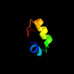
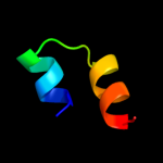
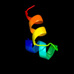
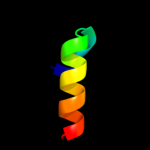
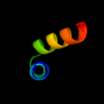
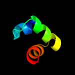
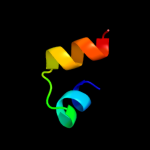
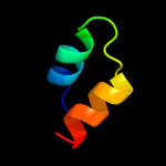
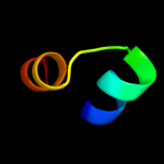
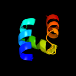
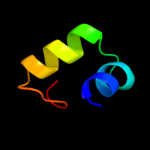
1 d1ifya_
76.4
23
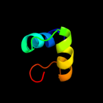
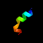
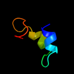
Fold: RuvA C-terminal domain-likeSuperfamily: UBA-likeFamily: UBA domain2 d1v92a_
60.7
24
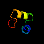
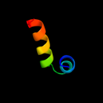
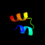
Fold: RuvA C-terminal domain-likeSuperfamily: UBA-likeFamily: TAP-C domain-like3 d1oqya1
53.1
23
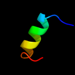
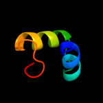
Fold: RuvA C-terminal domain-likeSuperfamily: UBA-likeFamily: UBA domain4 d2g3qa1
43.0
44
Fold: RuvA C-terminal domain-likeSuperfamily: UBA-likeFamily: UBA domain5 d1i9sa_
25.1
14
Fold: (Phosphotyrosine protein) phosphatases IISuperfamily: (Phosphotyrosine protein) phosphatases IIFamily: Dual specificity phosphatase-like6 c2gwoC_
22.7
32
PDB header: hydrolaseChain: C: PDB Molecule: dual specificity protein phosphatase 13;PDBTitle: crystal structure of tmdp
7 c2fw9A_
22.3
17
PDB header: lyaseChain: A: PDB Molecule: n5-carboxyaminoimidazole ribonucleotide mutase;PDBTitle: structure of pure (n5-carboxyaminoimidazole ribonucleotide mutase)2 h59f from the acidophilic bacterium acetobacter aceti, at ph 8
8 d1u11a_
21.9
17
Fold: Flavodoxin-likeSuperfamily: N5-CAIR mutase (phosphoribosylaminoimidazole carboxylase, PurE)Family: N5-CAIR mutase (phosphoribosylaminoimidazole carboxylase, PurE)9 d1wiva_
20.8
24
Fold: RuvA C-terminal domain-likeSuperfamily: UBA-likeFamily: UBA domain10 c3echC_
20.4
47
PDB header: transcription, transcription regulationChain: C: PDB Molecule: 25-mer fragment of protein armr;PDBTitle: the marr-family repressor mexr in complex with its antirepressor armr
11 c3trhI_
18.5
30
PDB header: lyaseChain: I: PDB Molecule: phosphoribosylaminoimidazole carboxylasePDBTitle: structure of a phosphoribosylaminoimidazole carboxylase catalytic2 subunit (pure) from coxiella burnetii
12 c3bq3A_
17.6
16
PDB header: cell cycle, ligaseChain: A: PDB Molecule: defective in cullin neddylation protein 1;PDBTitle: crystal structure of s. cerevisiae dcn1
13 c1yn9B_
16.9
32
PDB header: hydrolaseChain: B: PDB Molecule: polynucleotide 5'-phosphatase;PDBTitle: crystal structure of baculovirus rna 5'-phosphatase2 complexed with phosphate
14 c2ekkA_
16.8
38
PDB header: protein bindingChain: A: PDB Molecule: uba domain from e3 ubiquitin-protein ligasePDBTitle: solution structure of ruh-074, a human uba domain
15 c3nmeA_
15.6
23
PDB header: hydrolaseChain: A: PDB Molecule: sex4 glucan phosphatase;PDBTitle: structure of a plant phosphatase
16 c2jp7A_
15.2
14
PDB header: translationChain: A: PDB Molecule: mrna export factor mex67;PDBTitle: nmr structure of the mex67 uba domain
17 c2dakA_
14.9
30
PDB header: hydrolaseChain: A: PDB Molecule: ubiquitin carboxyl-terminal hydrolase 5;PDBTitle: solution structure of the second uba domain in the human2 ubiquitin specific protease 5 (isopeptidase 5)
18 d2dkla1
14.7
37
Fold: RuvA C-terminal domain-likeSuperfamily: UBA-likeFamily: UBA domain19 d1oqya2
13.9
20
Fold: RuvA C-terminal domain-likeSuperfamily: UBA-likeFamily: UBA domain20 c2esbA_
12.9
23
PDB header: hydrolaseChain: A: PDB Molecule: dual specificity protein phosphatase 18;PDBTitle: crystal structure of human dusp18
21 c3lp6D_
not modelled
12.5
27
PDB header: lyaseChain: D: PDB Molecule: phosphoribosylaminoimidazole carboxylase catalytic subunit;PDBTitle: crystal structure of rv3275c-e60a from mycobacterium tuberculosis at2 1.7a resolution
22 c2y96A_
not modelled
12.3
35
PDB header: hydrolaseChain: A: PDB Molecule: dual specificity phosphatase dupd1;PDBTitle: structure of human dual-specificity phosphatase 27
23 c2ywxA_
not modelled
11.9
18
PDB header: lyaseChain: A: PDB Molecule: phosphoribosylaminoimidazole carboxylase catalytic subunit;PDBTitle: crystal structure of phosphoribosylaminoimidazole carboxylase2 catalytic subunit from methanocaldococcus jannaschii
24 c2imgA_
not modelled
11.4
23
PDB header: hydrolaseChain: A: PDB Molecule: dual specificity protein phosphatase 23;PDBTitle: crystal structure of dual specificity protein phosphatase2 23 from homo sapiens in complex with ligand malate ion
25 c2c46B_
not modelled
11.4
13
PDB header: transferaseChain: B: PDB Molecule: mrna capping enzyme;PDBTitle: crystal structure of the human rna guanylyltransferase and2 5'-phosphatase
26 d1qcza_
not modelled
11.0
30
Fold: Flavodoxin-likeSuperfamily: N5-CAIR mutase (phosphoribosylaminoimidazole carboxylase, PurE)Family: N5-CAIR mutase (phosphoribosylaminoimidazole carboxylase, PurE)27 d1o4va_
not modelled
10.8
9
Fold: Flavodoxin-likeSuperfamily: N5-CAIR mutase (phosphoribosylaminoimidazole carboxylase, PurE)Family: N5-CAIR mutase (phosphoribosylaminoimidazole carboxylase, PurE)28 c2r0bA_
not modelled
10.4
27
PDB header: hydrolaseChain: A: PDB Molecule: serine/threonine/tyrosine-interacting protein;PDBTitle: crystal structure of human tyrosine phosphatase-like2 serine/threonine/tyrosine-interacting protein
29 c3orsD_
not modelled
10.4
18
PDB header: isomerase,biosynthetic proteinChain: D: PDB Molecule: n5-carboxyaminoimidazole ribonucleotide mutase;PDBTitle: crystal structure of n5-carboxyaminoimidazole ribonucleotide mutase2 from staphylococcus aureus
30 d1xmpa_
not modelled
10.1
30
Fold: Flavodoxin-likeSuperfamily: N5-CAIR mutase (phosphoribosylaminoimidazole carboxylase, PurE)Family: N5-CAIR mutase (phosphoribosylaminoimidazole carboxylase, PurE)31 d1oaia_
not modelled
9.5
23
Fold: RuvA C-terminal domain-likeSuperfamily: UBA-likeFamily: TAP-C domain-like32 c2h31A_
not modelled
9.5
22
PDB header: ligase, lyaseChain: A: PDB Molecule: multifunctional protein ade2;PDBTitle: crystal structure of human paics, a bifunctional carboxylase and2 synthetase in purine biosynthesis
33 c3emuA_
not modelled
9.5
18
PDB header: hydrolaseChain: A: PDB Molecule: leucine rich repeat and phosphatase domainPDBTitle: crystal structure of a leucine rich repeat and phosphatase2 domain containing protein from entamoeba histolytica
34 c3rggD_
not modelled
9.2
9
PDB header: lyaseChain: D: PDB Molecule: phosphoribosylaminoimidazole carboxylase, pure protein;PDBTitle: crystal structure of treponema denticola pure bound to air
35 c2hcmA_
not modelled
9.0
32
PDB header: hydrolaseChain: A: PDB Molecule: dual specificity protein phosphatase;PDBTitle: crystal structure of mouse putative dual specificity phosphatase2 complexed with zinc tungstate, new york structural genomics3 consortium
36 c3pcsB_
not modelled
8.5
36
PDB header: protein transport/transferaseChain: B: PDB Molecule: espg;PDBTitle: structure of espg-pak2 autoinhibitory ialpha3 helix complex
37 d1vega_
not modelled
7.9
27
Fold: RuvA C-terminal domain-likeSuperfamily: UBA-likeFamily: UBA domain38 c2crnA_
not modelled
7.8
24
PDB header: immune systemChain: A: PDB Molecule: ubash3a protein;PDBTitle: solution structure of the uba domain of human ubash3a2 protein
39 d1go5a_
not modelled
7.7
22
Fold: RuvA C-terminal domain-likeSuperfamily: UBA-likeFamily: TAP-C domain-like40 d1gyza_
not modelled
7.3
21
Fold: PABP domain-likeSuperfamily: Ribosomal protein L20Family: Ribosomal protein L2041 c1tr8A_
not modelled
7.2
23
PDB header: chaperoneChain: A: PDB Molecule: conserved protein (mth177);PDBTitle: crystal structure of archaeal nascent polypeptide-associated complex2 (aenac)
42 d1mkpa_
not modelled
6.9
27
Fold: (Phosphotyrosine protein) phosphatases IISuperfamily: (Phosphotyrosine protein) phosphatases IIFamily: Dual specificity phosphatase-like43 c2daiA_
not modelled
6.5
24
PDB header: structural genomics, unknown functionChain: A: PDB Molecule: ubiquitin associated domain containing 1;PDBTitle: solution structure of the first uba domain in the human2 ubiquitin associated domain containing 1 (ubadc1)
44 c1wrmA_
not modelled
6.5
23
PDB header: hydrolaseChain: A: PDB Molecule: dual specificity phosphatase 22;PDBTitle: crystal structure of jsp-1
45 d1wj7a1
not modelled
6.4
32
Fold: RuvA C-terminal domain-likeSuperfamily: UBA-likeFamily: UBA domain46 c2e0tA_
not modelled
6.3
27
PDB header: hydrolaseChain: A: PDB Molecule: dual specificity phosphatase 26;PDBTitle: crystal structure of catalytic domain of dual specificity phosphatase2 26, ms0830 from homo sapiens
47 d1veka_
not modelled
6.0
24
Fold: RuvA C-terminal domain-likeSuperfamily: UBA-likeFamily: UBA domain48 c3s4oB_
not modelled
6.0
18
PDB header: structural genomics, unknown functionChain: B: PDB Molecule: protein tyrosine phosphatase-like protein;PDBTitle: protein tyrosine phosphatase (putative) from leishmania major
49 d1pk1c1
not modelled
5.8
16
Fold: SAM domain-likeSuperfamily: SAM/Pointed domainFamily: SAM (sterile alpha motif) domain50 d1xb2b1
not modelled
5.7
26
Fold: RuvA C-terminal domain-likeSuperfamily: UBA-likeFamily: TS-N domain51 d1h2vc1
not modelled
5.7
27
Fold: alpha-alpha superhelixSuperfamily: ARM repeatFamily: MIF4G domain-like52 d1uqva_
not modelled
5.5
22
Fold: SAM domain-likeSuperfamily: SAM/Pointed domainFamily: SAM (sterile alpha motif) domain53 d1wuua2
not modelled
5.3
46
Fold: Ferredoxin-likeSuperfamily: GHMP Kinase, C-terminal domainFamily: Galactokinase


