Phyre Home Retrieve Phyre Job Id
Emaill.a.kelley@imperial.ac.uk
DescriptionP0AF61
DateWed Jan 25 15:20:33 GMT 2012
Unique Job ID0f5c145a1d7702fa

Summary 

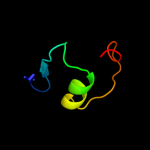
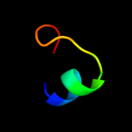
Top model
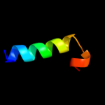
Image coloured by rainbow N → C terminus
Model (left) based on template d2rk5a1
Top template information
Fold:FAD-binding/transporter-associated domain-like
Superfamily:FAD-binding/transporter-associated domain-like
Family:CorC/HlyC domain-like
Confidence and coverage
Confidence: 18.6% Coverage: 34%
33 residues ( 34% of your sequence) have been modelled with 18.6% confidence by the single highest scoring template.
You may wish to submit your sequence to Phyrealarm. This will automatically scan your sequence every week for new potential templates as they appear in the Phyre2 library.
Please note: You must be registered and logged in to use Phyrealarm.
3D viewing
Interactive 3D view in Jmol

Sequence analysis 

Secondary structure and disorder prediction 

   1........10.........20.........30.........40.........50.........60
Sequence  MEGKNKFNTYVVSFDYPSSYSSVFLRLRSLMYDMNFSSIVADEYGIPRQLNENSFAITTS
Secondary structure 


SS confidence 


Disorder  ?????
?


?


Disorder confidence 


   .........70.........80.........90........
Sequence  LAASEIEDLIRLKCLDLPDIDFDLNIMTVDDYFRQFYK
Secondary structure 


SS confidence 


Disorder  ??


??????


??
Disorder confidence 


Confidence Key
High(9)                    Low (0)
?Disordered
Alpha helix
Beta strand

Domain analysis 

Hover over an aligned region to see model and summary info

Please note, only up to the top 20 hits are modelled to reduce computer load

RankAligned region

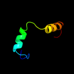
PDB 2rk5 chain A domain 1

3D model

Region: 48 - 83
Aligned: 33
Modelled: 36
Confidence: 18.6%
Identity: 15%
Fold: FAD-binding/transporter-associated domain-like
Superfamily: FAD-binding/transporter-associated domain-like
Family: CorC/HlyC domain-like

Phyre2

PDB 1t2s chain A

3D model

Region: 87 - 98
Aligned: 11
Modelled: 12
Confidence: 12.7%
Identity: 27%
Fold: SH3-like barrel
Superfamily: PAZ domain
Family: PAZ domain

Phyre2

PDB 1r6z chain A domain 1

3D model

Region: 87 - 98
Aligned: 12
Modelled: 12
Confidence: 11.2%
Identity: 25%
Fold: SH3-like barrel
Superfamily: PAZ domain
Family: PAZ domain

Phyre2

PDB 2be6 chain F

3D model

Region: 89 - 96
Aligned: 8
Modelled: 8
Confidence: 9.8%
Identity: 50%
PDB header:membrane protein
Chain: F: PDB Molecule:voltage-dependent l-type calcium channel alpha-1c subunit;
PDBTitle: 2.0 a crystal structure of the cav1.2 iq domain-ca/cam complex

Phyre2

PDB 3fqi chain A

3D model

Region: 71 - 85
Aligned: 15
Modelled: 15
Confidence: 9.6%
Identity: 40%
PDB header:protein binding
Chain: A: PDB Molecule:protein dom3z;
PDBTitle: crystal structure of the mouse dom3z

Phyre2

PDB 1nw3 chain A

3D model

Region: 62 - 82
Aligned: 21
Modelled: 21
Confidence: 9.0%
Identity: 43%
Fold: S-adenosyl-L-methionine-dependent methyltransferases
Superfamily: S-adenosyl-L-methionine-dependent methyltransferases
Family: Catalytic, N-terminal domain of histone methyltransferase Dot1l

Phyre2

PDB 1nw3 chain A

3D model

Region: 62 - 82
Aligned: 21
Modelled: 21
Confidence: 9.0%
Identity: 43%
PDB header:transferase
Chain: A: PDB Molecule:histone methyltransferase dot1l;
PDBTitle: structure of the catalytic domain of human dot1l, a non-set2 domain nucleosomal histone methyltransferase

Phyre2

PDB 1si2 chain A

3D model

Region: 87 - 98
Aligned: 12
Modelled: 12
Confidence: 8.1%
Identity: 50%
Fold: SH3-like barrel
Superfamily: PAZ domain
Family: PAZ domain

Phyre2

PDB 1h6z chain A domain 2

3D model

Region: 34 - 47
Aligned: 14
Modelled: 14
Confidence: 7.8%
Identity: 21%
Fold: The "swivelling" beta/beta/alpha domain
Superfamily: Phosphohistidine domain
Family: Pyruvate phosphate dikinase, central domain

Phyre2

PDB 2b0l chain A domain 1

3D model

Region: 37 - 48
Aligned: 12
Modelled: 12
Confidence: 7.5%
Identity: 50%
Fold: DNA/RNA-binding 3-helical bundle
Superfamily: "Winged helix" DNA-binding domain
Family: CodY HTH domain

Phyre2

PDB 2k9q chain B

3D model

Region: 40 - 77
Aligned: 38
Modelled: 38
Confidence: 7.5%
Identity: 34%
PDB header:structural genomics, unknown function
Chain: B: PDB Molecule:uncharacterized protein;
PDBTitle: solution nmr structure of hth_xre family transcriptional2 regulator bt_p548217 from bacteroides thetaiotaomicron.3 northeast structural genomics consortium target btr244.

Phyre2

PDB 3m1c chain B

3D model

Region: 60 - 78
Aligned: 19
Modelled: 19
Confidence: 7.5%
Identity: 37%
PDB header:viral protein
Chain: B: PDB Molecule:envelope glycoprotein l;
PDBTitle: crystal structure of the conserved herpesvirus fusion regulator2 complex gh-gl

Phyre2

PDB 1bw6 chain A

3D model

Region: 40 - 48
Aligned: 9
Modelled: 9
Confidence: 7.3%
Identity: 33%
Fold: DNA/RNA-binding 3-helical bundle
Superfamily: Homeodomain-like
Family: Centromere-binding

Phyre2

PDB 1hlv chain A domain 1

3D model

Region: 40 - 49
Aligned: 10
Modelled: 10
Confidence: 7.3%
Identity: 30%
Fold: DNA/RNA-binding 3-helical bundle
Superfamily: Homeodomain-like
Family: Centromere-binding

Phyre2

PDB 1kbl chain A domain 2

3D model

Region: 34 - 47
Aligned: 14
Modelled: 14
Confidence: 7.3%
Identity: 21%
Fold: The "swivelling" beta/beta/alpha domain
Superfamily: Phosphohistidine domain
Family: Pyruvate phosphate dikinase, central domain

Phyre2

PDB 2fc7 chain A domain 1

3D model

Region: 70 - 84
Aligned: 15
Modelled: 15
Confidence: 7.3%
Identity: 33%
Fold: RING/U-box
Superfamily: RING/U-box
Family: ZZ domain

Phyre2

PDB 3kow chain H

3D model

Region: 28 - 41
Aligned: 14
Modelled: 14
Confidence: 7.3%
Identity: 50%
PDB header:metal binding protein
Chain: H: PDB Molecule:d-ornithine aminomutase s component;
PDBTitle: crystal structure of ornithine 4,5 aminomutase backsoaked complex

Phyre2

PDB 3zrg chain B

3D model

Region: 87 - 92
Aligned: 6
Modelled: 6
Confidence: 7.3%
Identity: 100%
PDB header:protein binding
Chain: B: PDB Molecule:pexrd2 family secreted rxlr effector peptide, putative;
PDBTitle: crystal structure of rxlr effector pexrd2 from phytophthora infestans

Phyre2

PDB 1q8i chain A domain 2

3D model

Region: 1 - 73
Aligned: 68
Modelled: 73
Confidence: 7.1%
Identity: 13%
Fold: DNA/RNA polymerases
Superfamily: DNA/RNA polymerases
Family: DNA polymerase I

Phyre2

PDB 1kke chain A domain 2

3D model

Region: 83 - 92
Aligned: 10
Modelled: 10
Confidence: 7.0%
Identity: 50%
Fold: Virus attachment protein globular domain
Superfamily: Virus attachment protein globular domain
Family: Reovirus attachment protein sigma 1 head domain

Phyre2
1

d2rk5a1
2

d1t2sa_
3

d1r6za1
4

c2be6F_
5

c3fqiA_
6

d1nw3a_
7

c1nw3A_
8

d1si2a_
9

d1h6za2
10

d2b0la1
11

c2k9qB_
12

c3m1cB_
13

d1bw6a_
14

d1hlva1
15

d1kbla2
16

d2fc7a1
17

c3kowH_
18

c3zrgB_
19

d1q8ia2
20

d1kkea2
21


22


23


24


25


26


27


28


Detailed template information 

#
Template Alignment Coverage3D Model Confidence
% i.d. Template Information
1d2rk5a1


18.6 15 Fold:FAD-binding/transporter-associated domain-like
Superfamily:FAD-binding/transporter-associated domain-like
Family:CorC/HlyC domain-like
2d1t2sa_


12.7 27 Fold:SH3-like barrel
Superfamily:PAZ domain
Family:PAZ domain
3d1r6za1


11.2 25 Fold:SH3-like barrel
Superfamily:PAZ domain
Family:PAZ domain
4c2be6F_


9.8 50 PDB header:membrane protein
Chain: F: PDB Molecule:voltage-dependent l-type calcium channel alpha-1c subunit;
PDBTitle: 2.0 a crystal structure of the cav1.2 iq domain-ca/cam complex
5c3fqiA_


9.6 40 PDB header:protein binding
Chain: A: PDB Molecule:protein dom3z;
PDBTitle: crystal structure of the mouse dom3z
6d1nw3a_


9.0 43 Fold:S-adenosyl-L-methionine-dependent methyltransferases
Superfamily:S-adenosyl-L-methionine-dependent methyltransferases
Family:Catalytic, N-terminal domain of histone methyltransferase Dot1l
7c1nw3A_


9.0 43 PDB header:transferase
Chain: A: PDB Molecule:histone methyltransferase dot1l;
PDBTitle: structure of the catalytic domain of human dot1l, a non-set2 domain nucleosomal histone methyltransferase
8d1si2a_


8.1 50 Fold:SH3-like barrel
Superfamily:PAZ domain
Family:PAZ domain
9d1h6za2


7.8 21 Fold:The "swivelling" beta/beta/alpha domain
Superfamily:Phosphohistidine domain
Family:Pyruvate phosphate dikinase, central domain
10d2b0la1


7.5 50 Fold:DNA/RNA-binding 3-helical bundle
Superfamily:"Winged helix" DNA-binding domain
Family:CodY HTH domain
11c2k9qB_


7.5 34 PDB header:structural genomics, unknown function
Chain: B: PDB Molecule:uncharacterized protein;
PDBTitle: solution nmr structure of hth_xre family transcriptional2 regulator bt_p548217 from bacteroides thetaiotaomicron.3 northeast structural genomics consortium target btr244.
12c3m1cB_


7.5 37 PDB header:viral protein
Chain: B: PDB Molecule:envelope glycoprotein l;
PDBTitle: crystal structure of the conserved herpesvirus fusion regulator2 complex gh-gl
13d1bw6a_


7.3 33 Fold:DNA/RNA-binding 3-helical bundle
Superfamily:Homeodomain-like
Family:Centromere-binding
14d1hlva1


7.3 30 Fold:DNA/RNA-binding 3-helical bundle
Superfamily:Homeodomain-like
Family:Centromere-binding
15d1kbla2


7.3 21 Fold:The "swivelling" beta/beta/alpha domain
Superfamily:Phosphohistidine domain
Family:Pyruvate phosphate dikinase, central domain
16d2fc7a1


7.3 33 Fold:RING/U-box
Superfamily:RING/U-box
Family:ZZ domain
17c3kowH_


7.3 50 PDB header:metal binding protein
Chain: H: PDB Molecule:d-ornithine aminomutase s component;
PDBTitle: crystal structure of ornithine 4,5 aminomutase backsoaked complex
18c3zrgB_


7.3 100 PDB header:protein binding
Chain: B: PDB Molecule:pexrd2 family secreted rxlr effector peptide, putative;
PDBTitle: crystal structure of rxlr effector pexrd2 from phytophthora infestans
19d1q8ia2


7.1 13 Fold:DNA/RNA polymerases
Superfamily:DNA/RNA polymerases
Family:DNA polymerase I
20d1kkea2


7.0 50 Fold:Virus attachment protein globular domain
Superfamily:Virus attachment protein globular domain
Family:Reovirus attachment protein sigma 1 head domain
21d2ghvc1


not modelled 6.9 25 Fold:SARS receptor-binding domain-like
Superfamily:SARS receptor-binding domain-like
Family:SARS receptor-binding domain-like
22c2elhA_


not modelled 6.4 33 PDB header:dna binding protein
Chain: A: PDB Molecule:cg11849-pa;
PDBTitle: solution structure of the cenp-b n-terminal dna-binding2 domain of fruit fly distal antenna cg11849-pa
23d2coba1


not modelled 6.1 44 Fold:DNA/RNA-binding 3-helical bundle
Superfamily:Homeodomain-like
Family:Psq domain
24d2dloa2


not modelled 5.9 29 Fold:Glucocorticoid receptor-like (DNA-binding domain)
Superfamily:Glucocorticoid receptor-like (DNA-binding domain)
Family:LIM domain
25d1s5ja2


not modelled 5.7 15 Fold:DNA/RNA polymerases
Superfamily:DNA/RNA polymerases
Family:DNA polymerase I
26d1vbga2


not modelled 5.7 25 Fold:The "swivelling" beta/beta/alpha domain
Superfamily:Phosphohistidine domain
Family:Pyruvate phosphate dikinase, central domain
27c1kkeA_


not modelled 5.3 50 PDB header:viral protein
Chain: A: PDB Molecule:sigma 1 protein;
PDBTitle: crystal structure of reovirus attachment protein sigma12 trimer
28c3o7vX_


not modelled 5.1 25 PDB header:rna binding protein/rna
Chain: X: PDB Molecule:piwi-like protein 1;
PDBTitle: crystal structure of human hiwi1 (v361m) paz domain (residues 277-399)2 in complex with 14-mer rna (12-bp + 2-nt overhang) containing 2'-och33 at its 3'-end

Binding site prediction 

Due to computational demand, binding site predictions are not run for batch jobs

If you want to predict binding sites, please manually submit your model of choice to 3DLigandSite


Phyre is for academic use only

Please cite: Protein structure prediction on the web: a case study using the Phyre server
Kelley LA and Sternberg MJE. Nature Protocols 4, 363 - 371 (2009) [pdf] [Import into BibTeX]
 
If you use the binding site predictions from 3DLigandSite, please also cite:
3DLigandSite: predicting ligand-binding sites using similar structures.
Wass MN, Kelley LA and Sternberg MJ Nucleic Acids Research 38, W469-73 (2010) [PubMed]
 
© Structural Bioinformatics Group
Imperial College London
Lawrence Kelley, Benjamin Jefferys
Disclaimer
Terms and Conditions
Component software
Template detection: HHpred 1.51
Secondary structure prediction: Psi-pred 2.5
Disorder prediction: Disopred 2.4
Transmembrane prediction: Memsat_SVM
Multi-template modelling and ab initio: Poing 1.0