Phyre Home Retrieve Phyre Job Id
Emaill.a.kelley@imperial.ac.uk
DescriptionP62395
DateThu Jan 5 12:07:29 GMT 2012
Unique Job ID0f3a5c7f0451b9f3

Summary 

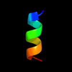
Top model
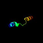
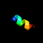
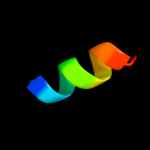
Image coloured by rainbow N → C terminus
Model (left) based on template c3ibpA_
Top template information
PDB header:cell cycle
Chain: A: PDB Molecule:chromosome partition protein mukb;
PDBTitle: the crystal structure of the dimerization domain of escherichia coli2 structural maintenance of chromosomes protein mukb
Confidence and coverage
Confidence: 24.8% Coverage: 19%
33 residues ( 19% of your sequence) have been modelled with 24.8% confidence by the single highest scoring template.
You may wish to submit your sequence to Phyrealarm. This will automatically scan your sequence every week for new potential templates as they appear in the Phyre2 library.
Please note: You must be registered and logged in to use Phyrealarm.
3D viewing
Interactive 3D view in Jmol

Sequence analysis 

Secondary structure and disorder prediction 

   1........10.........20.........30.........40.........50.........60
Sequence  MSGILTRWRQFGKRYFWPHLLLGMVAASLGLPALSNAAEPNAPAKATTRNHEPSAKVNFG
Secondary structure 


SS confidence 


Disorder  ????


??????????????


Disorder confidence 


   .........70.........80.........90.........100.........110.........120
Sequence  QLALLEANTRRPNSNYSVDYWHQHAIRTVIRHLSFAMAPQTLPVAEESLPLQAQHLALLD
Secondary structure 


SS confidence 


Disorder 


?????????
?


????


????????


Disorder confidence 


   .........130.........140.........150.........160.........170
Sequence  TLSALLTQEGTPSEKGYRIDYAHFTPQAKFSTPVWISQAQGIRAGPQRLT
Secondary structure 


SS confidence 


Disorder 


???????????????????????

?


?


?
???????
Disorder confidence 


Confidence Key
High(9)                    Low (0)
?Disordered
Alpha helix
Beta strand

Domain analysis 

Hover over an aligned region to see model and summary info

Please note, only up to the top 20 hits are modelled to reduce computer load

RankAligned region

PDB 3ibp chain A

3D model

Region: 82 - 114
Aligned: 33
Modelled: 33
Confidence: 24.8%
Identity: 18%
PDB header:cell cycle
Chain: A: PDB Molecule:chromosome partition protein mukb;
PDBTitle: the crystal structure of the dimerization domain of escherichia coli2 structural maintenance of chromosomes protein mukb

Phyre2

PDB 3ost chain A

3D model

Region: 4 - 12
Aligned: 9
Modelled: 9
Confidence: 20.8%
Identity: 44%
PDB header:lipid binding protein
Chain: A: PDB Molecule:serine/threonine-protein kinase kcc4;
PDBTitle: structure of the kinase associated-1 (ka1) from kcc4p

Phyre2

PDB 1wdk chain A domain 2

3D model

Region: 4 - 30
Aligned: 27
Modelled: 27
Confidence: 12.8%
Identity: 15%
Fold: 6-phosphogluconate dehydrogenase C-terminal domain-like
Superfamily: 6-phosphogluconate dehydrogenase C-terminal domain-like
Family: HCDH C-domain-like

Phyre2

PDB 1jb0 chain X

3D model

Region: 14 - 24
Aligned: 11
Modelled: 11
Confidence: 12.4%
Identity: 55%
PDB header:photosynthesis
Chain: X: PDB Molecule:photosystem i subunit psax;
PDBTitle: crystal structure of photosystem i: a photosynthetic reaction center2 and core antenna system from cyanobacteria

Phyre2

PDB 1jb0 chain X

3D model

Region: 14 - 24
Aligned: 11
Modelled: 11
Confidence: 12.4%
Identity: 55%
Fold: Single transmembrane helix
Superfamily: Subunit PsaX of photosystem I reaction centre
Family: Subunit PsaX of photosystem I reaction centre

Phyre2

PDB 3pcq chain X

3D model

Region: 14 - 24
Aligned: 11
Modelled: 11
Confidence: 11.6%
Identity: 55%
PDB header:photosynthesis
Chain: X: PDB Molecule:photosystem i 4.8k protein;
PDBTitle: femtosecond x-ray protein nanocrystallography

Phyre2

PDB 1zmb chain A domain 1

3D model

Region: 9 - 17
Aligned: 9
Modelled: 9
Confidence: 11.0%
Identity: 67%
Fold: Flavodoxin-like
Superfamily: SGNH hydrolase
Family: Putative acetylxylan esterase-like

Phyre2

PDB 1yle chain A domain 1

3D model

Region: 1 - 16
Aligned: 16
Modelled: 16
Confidence: 10.3%
Identity: 25%
Fold: Acyl-CoA N-acyltransferases (Nat)
Superfamily: Acyl-CoA N-acyltransferases (Nat)
Family: AstA-like

Phyre2

PDB 2ks1 chain B

3D model

Region: 20 - 31
Aligned: 12
Modelled: 12
Confidence: 9.5%
Identity: 58%
PDB header:transferase
Chain: B: PDB Molecule:epidermal growth factor receptor;
PDBTitle: heterodimeric association of transmembrane domains of erbb1 and erbb22 receptors enabling kinase activation

Phyre2

PDB 2fiq chain A domain 1

3D model

Region: 12 - 19
Aligned: 8
Modelled: 8
Confidence: 9.4%
Identity: 63%
Fold: TIM beta/alpha-barrel
Superfamily: Aldolase
Family: GatZ-like

Phyre2

PDB 2izv chain A domain 1

3D model

Region: 83 - 94
Aligned: 12
Modelled: 12
Confidence: 9.2%
Identity: 50%
Fold: SOCS box-like
Superfamily: SOCS box-like
Family: SOCS box-like

Phyre2

PDB 3nnq chain A

3D model

Region: 3 - 20
Aligned: 18
Modelled: 12
Confidence: 8.2%
Identity: 6%
PDB header:viral protein
Chain: A: PDB Molecule:n-terminal domain of moloney murine leukemia virus
PDBTitle: crystal structure of the n-terminal domain of moloney murine leukemia2 virus integrase, northeast structural genomics consortium target or3

Phyre2

PDB 1hly chain A

3D model

Region: 161 - 165
Aligned: 5
Modelled: 5
Confidence: 7.9%
Identity: 100%
Fold: Knottins (small inhibitors, toxins, lectins)
Superfamily: Scorpion toxin-like
Family: Short-chain scorpion toxins

Phyre2

PDB 1g3n chain C domain 2

3D model

Region: 75 - 90
Aligned: 15
Modelled: 16
Confidence: 7.8%
Identity: 40%
Fold: Cyclin-like
Superfamily: Cyclin-like
Family: Cyclin

Phyre2

PDB 1vf5 chain G

3D model

Region: 20 - 30
Aligned: 11
Modelled: 11
Confidence: 7.0%
Identity: 64%
Fold: Single transmembrane helix
Superfamily: PetG subunit of the cytochrome b6f complex
Family: PetG subunit of the cytochrome b6f complex

Phyre2

PDB 1vf5 chain G

3D model

Region: 20 - 30
Aligned: 11
Modelled: 11
Confidence: 7.0%
Identity: 64%
PDB header:photosynthesis
Chain: G: PDB Molecule:protein pet g;
PDBTitle: crystal structure of cytochrome b6f complex from m.laminosus

Phyre2

PDB 2e74 chain G domain 1

3D model

Region: 20 - 30
Aligned: 11
Modelled: 11
Confidence: 6.9%
Identity: 64%
Fold: Single transmembrane helix
Superfamily: PetG subunit of the cytochrome b6f complex
Family: PetG subunit of the cytochrome b6f complex

Phyre2

PDB 2e76 chain D

3D model

Region: 11 - 30
Aligned: 20
Modelled: 20
Confidence: 6.7%
Identity: 30%
PDB header:photosynthesis
Chain: D: PDB Molecule:cytochrome b6-f complex iron-sulfur subunit;
PDBTitle: crystal structure of the cytochrome b6f complex with tridecyl-2 stigmatellin (tds) from m.laminosus

Phyre2

PDB 2qdo chain C

3D model

Region: 88 - 93
Aligned: 6
Modelled: 6
Confidence: 6.5%
Identity: 67%
PDB header:photosynthesis
Chain: C: PDB Molecule:nbla protein;
PDBTitle: nbla protein from t. vulcanus

Phyre2

PDB 3d7v chain B

3D model

Region: 151 - 158
Aligned: 8
Modelled: 8
Confidence: 6.2%
Identity: 38%
PDB header:apoptosis
Chain: B: PDB Molecule:bcl-2-like protein 11;
PDBTitle: crystal structure of mcl-1 in complex with an mcl-12 selective bh3 ligand

Phyre2
1

c3ibpA_
2

c3ostA_
3

d1wdka2
4

c1jb0X_
5

d1jb0x_
6

c3pcqX_
7

d1zmba1
8

d1ylea1
9

c2ks1B_
10

d2fiqa1
11

d2izva1
12

c3nnqA_
13

d1hlya_
14

d1g3nc2
15

d1vf5g_
16

c1vf5G_
17

d2e74g1
18

c2e76D_
19

c2qdoC_
20

c3d7vB_
21


22


23


24


25


26


27


28


29


30


31


Detailed template information 

#
Template Alignment Coverage3D Model Confidence
% i.d. Template Information
1c3ibpA_


24.8 18 PDB header:cell cycle
Chain: A: PDB Molecule:chromosome partition protein mukb;
PDBTitle: the crystal structure of the dimerization domain of escherichia coli2 structural maintenance of chromosomes protein mukb
2c3ostA_


20.8 44 PDB header:lipid binding protein
Chain: A: PDB Molecule:serine/threonine-protein kinase kcc4;
PDBTitle: structure of the kinase associated-1 (ka1) from kcc4p
3d1wdka2


12.8 15 Fold:6-phosphogluconate dehydrogenase C-terminal domain-like
Superfamily:6-phosphogluconate dehydrogenase C-terminal domain-like
Family:HCDH C-domain-like
4c1jb0X_


12.4 55 PDB header:photosynthesis
Chain: X: PDB Molecule:photosystem i subunit psax;
PDBTitle: crystal structure of photosystem i: a photosynthetic reaction center2 and core antenna system from cyanobacteria
5d1jb0x_


12.4 55 Fold:Single transmembrane helix
Superfamily:Subunit PsaX of photosystem I reaction centre
Family:Subunit PsaX of photosystem I reaction centre
6c3pcqX_


11.6 55 PDB header:photosynthesis
Chain: X: PDB Molecule:photosystem i 4.8k protein;
PDBTitle: femtosecond x-ray protein nanocrystallography
7d1zmba1


11.0 67 Fold:Flavodoxin-like
Superfamily:SGNH hydrolase
Family:Putative acetylxylan esterase-like
8d1ylea1


10.3 25 Fold:Acyl-CoA N-acyltransferases (Nat)
Superfamily:Acyl-CoA N-acyltransferases (Nat)
Family:AstA-like
9c2ks1B_


9.5 58 PDB header:transferase
Chain: B: PDB Molecule:epidermal growth factor receptor;
PDBTitle: heterodimeric association of transmembrane domains of erbb1 and erbb22 receptors enabling kinase activation
10d2fiqa1


9.4 63 Fold:TIM beta/alpha-barrel
Superfamily:Aldolase
Family:GatZ-like
11d2izva1


9.2 50 Fold:SOCS box-like
Superfamily:SOCS box-like
Family:SOCS box-like
12c3nnqA_


8.2 6 PDB header:viral protein
Chain: A: PDB Molecule:n-terminal domain of moloney murine leukemia virus
PDBTitle: crystal structure of the n-terminal domain of moloney murine leukemia2 virus integrase, northeast structural genomics consortium target or3
13d1hlya_


7.9 100 Fold:Knottins (small inhibitors, toxins, lectins)
Superfamily:Scorpion toxin-like
Family:Short-chain scorpion toxins
14d1g3nc2


7.8 40 Fold:Cyclin-like
Superfamily:Cyclin-like
Family:Cyclin
15d1vf5g_


7.0 64 Fold:Single transmembrane helix
Superfamily:PetG subunit of the cytochrome b6f complex
Family:PetG subunit of the cytochrome b6f complex
16c1vf5G_


7.0 64 PDB header:photosynthesis
Chain: G: PDB Molecule:protein pet g;
PDBTitle: crystal structure of cytochrome b6f complex from m.laminosus
17d2e74g1


6.9 64 Fold:Single transmembrane helix
Superfamily:PetG subunit of the cytochrome b6f complex
Family:PetG subunit of the cytochrome b6f complex
18c2e76D_


6.7 30 PDB header:photosynthesis
Chain: D: PDB Molecule:cytochrome b6-f complex iron-sulfur subunit;
PDBTitle: crystal structure of the cytochrome b6f complex with tridecyl-2 stigmatellin (tds) from m.laminosus
19c2qdoC_


6.5 67 PDB header:photosynthesis
Chain: C: PDB Molecule:nbla protein;
PDBTitle: nbla protein from t. vulcanus
20c3d7vB_


6.2 38 PDB header:apoptosis
Chain: B: PDB Molecule:bcl-2-like protein 11;
PDBTitle: crystal structure of mcl-1 in complex with an mcl-12 selective bh3 ligand
21c2kfdA_


not modelled 6.1 50 PDB header:nuclear protein
Chain: A: PDB Molecule:pre-mrna-processing protein prp40;
PDBTitle: prp40 ff4 domain
22c1bm4A_


not modelled 6.0 40 PDB header:viral protein
Chain: A: PDB Molecule:protein (moloney murine leukemia virus capsid);
PDBTitle: momlv capsid protein major homology region peptide analog
23c2vy8A_


not modelled 6.0 43 PDB header:transcription
Chain: A: PDB Molecule:polymerase basic protein 2;
PDBTitle: the 627-domain from influenza a virus polymerase pb22 subunit with glu-627
24c3io9B_


not modelled 6.0 38 PDB header:apoptosis
Chain: B: PDB Molecule:bcl-2-like protein 11;
PDBTitle: biml12y in complex with mcl-1
25c3kj2B_


not modelled 6.0 38 PDB header:apoptosis
Chain: B: PDB Molecule:bcl-2-like protein 11;
PDBTitle: mcl-1 in complex with bim bh3 mutant f4ae
26c2nl9B_


not modelled 5.9 38 PDB header:apoptosis
Chain: B: PDB Molecule:bcl-2-like protein 11;
PDBTitle: crystal structure of the mcl-1:bim bh3 complex
27d2nysa1


not modelled 5.9 54 Fold:SspB-like
Superfamily:SspB-like
Family:AGR C 3712p-like
28c2nysA_


not modelled 5.9 54 PDB header:structural genomics, unknown function
Chain: A: PDB Molecule:agr_c_3712p;
PDBTitle: x-ray crystal structure of protein agr_c_3712 from2 agrobacterium tumefaciens. northeast structural genomics3 consortium target atr88.
29c3rfrI_


not modelled 5.6 35 PDB header:oxidoreductase
Chain: I: PDB Molecule:pmob;
PDBTitle: crystal structure of particulate methane monooxygenase (pmmo) from2 methylocystis sp. strain m
30c2qazC_


not modelled 5.3 31 PDB header:hydrolase activator
Chain: C: PDB Molecule:sspb protein;
PDBTitle: structure of c. crescentus sspb ortholog
31d2e74b1


not modelled 5.1 36 Fold:a domain/subunit of cytochrome bc1 complex (Ubiquinol-cytochrome c reductase)
Superfamily:a domain/subunit of cytochrome bc1 complex (Ubiquinol-cytochrome c reductase)
Family:a domain/subunit of cytochrome bc1 complex (Ubiquinol-cytochrome c reductase)

Binding site prediction 

Due to computational demand, binding site predictions are not run for batch jobs

If you want to predict binding sites, please manually submit your model of choice to 3DLigandSite


Phyre is for academic use only

Please cite: Protein structure prediction on the web: a case study using the Phyre server
Kelley LA and Sternberg MJE. Nature Protocols 4, 363 - 371 (2009) [pdf] [Import into BibTeX]
 
If you use the binding site predictions from 3DLigandSite, please also cite:
3DLigandSite: predicting ligand-binding sites using similar structures.
Wass MN, Kelley LA and Sternberg MJ Nucleic Acids Research 38, W469-73 (2010) [PubMed]
 
© Structural Bioinformatics Group
Imperial College London
Lawrence Kelley, Benjamin Jefferys
Disclaimer
Terms and Conditions
Component software
Template detection: HHpred 1.51
Secondary structure prediction: Psi-pred 2.5
Disorder prediction: Disopred 2.4
Transmembrane prediction: Memsat_SVM
Multi-template modelling and ab initio: Poing 1.0