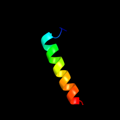
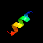
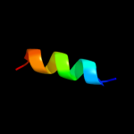
1 c2kmfA_
30.8
25
PDB header: photosynthesisChain: A: PDB Molecule: photosystem ii 11 kda protein;PDBTitle: solution structure of psb27 from cyanobacterial photosystem2 ii
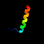
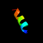
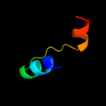
2 c2vdaB_
27.7
36
PDB header: protein transportChain: B: PDB Molecule: maltoporin;PDBTitle: solution structure of the seca-signal peptide complex
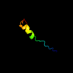
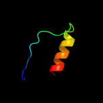
3 d1w6ua_
21.8
22
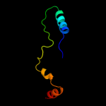
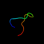
Fold: NAD(P)-binding Rossmann-fold domainsSuperfamily: NAD(P)-binding Rossmann-fold domainsFamily: Tyrosine-dependent oxidoreductases4 c1p5lA_
21.4
54
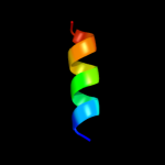
PDB header: ribosomeChain: A: PDB Molecule: 19-mer peptide from 50s ribosomal protein l1;PDBTitle: hp (2-20) substitution phe5 to ser modification in sds-d252 micelles
5 c1p5kA_
20.0
54
PDB header: ribosomeChain: A: PDB Molecule: 19-mer peptide from 50s ribosomal protein l1;PDBTitle: hp (2-20) substitution ser to leu11 modification in sds-d252 micelles
6 d1txka2
14.3
50
Fold: SupersandwichSuperfamily: Galactose mutarotase-likeFamily: MdoG-like7 c1txkA_
13.2
50
PDB header: biosynthetic proteinChain: A: PDB Molecule: glucans biosynthesis protein g;PDBTitle: crystal structure of escherichia coli opgg
8 c1p0oA_
13.0
54
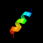
PDB header: ribosomeChain: A: PDB Molecule: 19-mer peptide from 50s ribosomal protein l1;PDBTitle: hp (2-20) substitution of trp for gln and asp at position2 17 and 19 modification in sds-d25 micelles
9 c1p0jA_
12.9
54
PDB header: ribosomeChain: A: PDB Molecule: 19-mer peptide from 50s ribosomal protein l1;PDBTitle: hp (2-20) substitution asp to trp modification in sds-d252 micelles
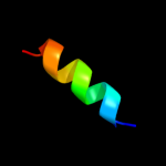
10 c1ot0A_
12.9
54
PDB header: antibioticChain: A: PDB Molecule: 50s ribosomal protein l1;PDBTitle: structure of antimicrobial peptide, hp (2-20) and its2 analogues derived from helicobacter pylori, as determined3 by 1h nmr spectroscopy
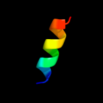
11 c1p0lA_
12.7
54
PDB header: ribosomeChain: A: PDB Molecule: 19-mer peptide from 50s ribosomal protein l1;PDBTitle: hp (2-20) substitution gln to trp modification in sds-d252 micelles
12 c1p0gA_
12.6
54
PDB header: ribosomeChain: A: PDB Molecule: 19-mer peptide from 50s ribosomal protein l1;PDBTitle: structure of antimicrobial peptide, hp (2-20) and its2 analogues derived from helicobacter pylori, as determined3 by 1h nmr spectroscopy
13 c3gwnA_
12.6
17
PDB header: oxidoreductaseChain: A: PDB Molecule: probable fad-linked sulfhydryl oxidase r596;PDBTitle: crystal structure of the fad binding domain from mimivirus sulfhydryl2 oxidase r596
14 c2yytA_
12.0
21
PDB header: lyaseChain: A: PDB Molecule: orotidine 5'-phosphate decarboxylase;PDBTitle: crystal structure of uncharacterized conserved protein from2 geobacillus kaustophilus
15 d1t95a1
10.2
27
Fold: RuvA C-terminal domain-likeSuperfamily: Hypothetical protein AF0491, middle domainFamily: Hypothetical protein AF0491, middle domain16 d1fcqa_
9.4
22
Fold: TIM beta/alpha-barrelSuperfamily: (Trans)glycosidasesFamily: Bee venom hyaluronidase17 c1fcuA_
9.3
22
PDB header: hydrolaseChain: A: PDB Molecule: hyaluronoglucosaminidase;PDBTitle: crystal structure (trigonal) of bee venom hyaluronidase
18 c3eabD_
8.6
38
PDB header: cell cycleChain: D: PDB Molecule: spastin;PDBTitle: crystal structure of spastin mit in complex with escrt iii
19 c2z02A_
7.8
29
PDB header: ligaseChain: A: PDB Molecule: phosphoribosylaminoimidazole-succinocarboxamidePDBTitle: crystal structure of2 phosphoribosylaminoimidazolesuccinocarboxamide synthase3 wit atp from methanocaldococcus jannaschii
20 d1bh9b_
7.5
26
Fold: Histone-foldSuperfamily: Histone-foldFamily: TBP-associated factors, TAFs21 c2h31A_
not modelled
7.0
22
PDB header: ligase, lyaseChain: A: PDB Molecule: multifunctional protein ade2;PDBTitle: crystal structure of human paics, a bifunctional carboxylase and2 synthetase in purine biosynthesis
22 c2dl1A_
not modelled
6.3
22
PDB header: protein transportChain: A: PDB Molecule: spartin;PDBTitle: solution structure of the mit domain from human spartin
23 d1cjca1
not modelled
5.9
12
Fold: FAD/NAD(P)-binding domainSuperfamily: FAD/NAD(P)-binding domainFamily: C-terminal domain of adrenodoxin reductase-like24 c3kreA_
not modelled
5.5
17
PDB header: ligaseChain: A: PDB Molecule: phosphoribosylaminoimidazole-succinocarboxamide synthase;PDBTitle: crystal structure of phosphoribosylaminoimidazole-succinocarboxamide2 synthase from ehrlichia chaffeensis at 1.8a resolution
25 d1bf2a2
not modelled
5.4
14
Fold: Glycosyl hydrolase domainSuperfamily: Glycosyl hydrolase domainFamily: alpha-Amylases, C-terminal beta-sheet domain26 c2x3bB_
not modelled
5.1
27
PDB header: hydrolaseChain: B: PDB Molecule: toxic extracellular endopeptidase;PDBTitle: asap1 inactive mutant e294a, an extracellular toxic zinc2 metalloendopeptidase