| Secondary structure and disorder prediction | |
| | |
1 | . | . | . | . | . | . | . | . | 10 | . | . | . | . | . | . | . | . | . | 20 | . | . | . | . | . | . | . | . | . | 30 | . | . | . | . | . | . | . | . | . | 40 | . |
| Sequence | |
M | M | K | R | L | I | V | L | V | L | L | A | S | T | L | L | T | G | C | N | T | A | R | G | F | G | E | D | I | K | H | L | G | N | S | I | S | R | A | A | S |
| Secondary structure | |
|  | | | | | | | | | | | | | | | | | | | | | | | | | | | | | | | | | | | | | |
|
|
| SS confidence | |
|
|
|
|
|
|
|
|
|
|
|
|
|
|
|
|
|
|
|
|
|
|
|
|
|
|
|
|
|
|
|
|
|
|
|
|
|
|
|
|
|
| Disorder | |
? |
|
|
|
|
|
|
|
|
|
|
|
|
|
|
|
|
|
|
|
|
|
|
|
|
|
|
|
|
|
| ? |
| ? |
|
|
| ? | ? | ? | ? |
| Disorder confidence | |
|
|
|
|
|
|
|
|
|
|
|
|
|
|
|
|
|
|
|
|
|
|
|
|
|
|
|
|
|
|
|
|
|
|
|
|
|
|
|
|
|
| |
| Confidence Key |
| High(9) | |
|
|
|
|
|
|
|
|
|
Low (0) |
| ? | Disordered |
| Alpha helix |
  | Beta strand |
Hover over an aligned region to see model and summary info
Please note, only up to the top 20 hits are modelled to reduce computer load
|
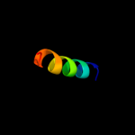
| 1 |
|
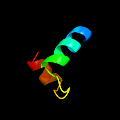
PDB 2rh3 chain A domain 1
Region: 1 - 29
Aligned: 29
Modelled: 29
Confidence: 25.9%
Identity: 24%
Fold: Ribbon-helix-helix
Superfamily: Ribbon-helix-helix
Family: VirC2-like
Phyre2
| 2 |
|
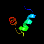
PDB 1pyv chain A
Region: 10 - 41
Aligned: 30
Modelled: 32
Confidence: 11.6%
Identity: 50%
PDB header:hydrolase
Chain: A: PDB Molecule:atp synthase beta chain, mitochondrial precursor;
PDBTitle: nmr solution structure of the mitochondrial f1b presequence2 peptide from nicotiana plumbaginifolia
Phyre2
| 3 |
|
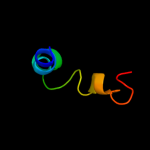
PDB 1sp8 chain A domain 2
Region: 18 - 38
Aligned: 21
Modelled: 21
Confidence: 9.0%
Identity: 38%
Fold: Glyoxalase/Bleomycin resistance protein/Dihydroxybiphenyl dioxygenase
Superfamily: Glyoxalase/Bleomycin resistance protein/Dihydroxybiphenyl dioxygenase
Family: Extradiol dioxygenases
Phyre2
| 4 |
|
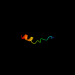
PDB 1deb chain A
Region: 20 - 36
Aligned: 17
Modelled: 17
Confidence: 7.4%
Identity: 35%
PDB header:structural protein
Chain: A: PDB Molecule:adenomatous polyposis coli protein;
PDBTitle: crystal structure of the n-terminal coiled coil domain from2 apc
Phyre2
| 5 |
|
PDB 1nh9 chain A
Region: 33 - 39
Aligned: 7
Modelled: 7
Confidence: 6.7%
Identity: 43%
Fold: IF3-like
Superfamily: AlbA-like
Family: DNA-binding protein AlbA
Phyre2
|
| Detailed template information | |
Due to computational demand, binding site predictions are not run for batch jobs
If you want to predict binding sites, please manually submit your model of choice to 3DLigandSite
Phyre is for academic use only
| Please cite: Protein structure prediction on
the web: a case study using the Phyre server |
| Kelley LA and Sternberg MJE. Nature Protocols
4, 363 - 371 (2009) [pdf] [Import into BibTeX] |
| |
| If you use the binding site
predictions from 3DLigandSite, please also cite: |
| 3DLigandSite: predicting ligand-binding sites using similar structures. |
| Wass MN, Kelley LA and Sternberg
MJ Nucleic Acids Research 38, W469-73 (2010) [PubMed] |
| |
|
|
|
|