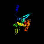
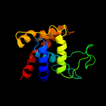
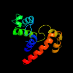
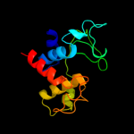
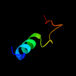
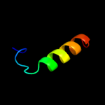
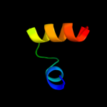
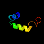
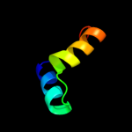
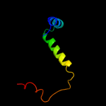
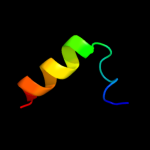
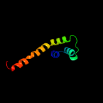
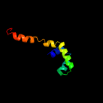
1 c3fi7A_
100.0
24
PDB header: hydrolaseChain: A: PDB Molecule: lmo1076 protein;PDBTitle: crystal structure of the autolysin auto (lmo1076) from listeria2 monocytogenes, catalytic domain
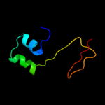
2 c2zycA_
99.9
24
PDB header: hydrolaseChain: A: PDB Molecule: peptidoglycan hydrolase flgj;PDBTitle: crystal structure of peptidoglycan hydrolase from2 sphingomonas sp. a1
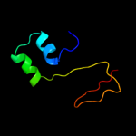
3 d1qsaa2
86.9
16
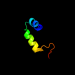
Fold: Lysozyme-likeSuperfamily: Lysozyme-likeFamily: Bacterial muramidase, catalytic domain4 c3gxkB_
71.0
15
PDB header: hydrolaseChain: B: PDB Molecule: goose-type lysozyme 1;PDBTitle: the crystal structure of g-type lysozyme from atlantic cod2 (gadus morhua l.) in complex with nag oligomers sheds new3 light on substrate binding and the catalytic mechanism.4 native structure to 1.9
5 d1gbsa_
48.7
15
Fold: Lysozyme-likeSuperfamily: Lysozyme-likeFamily: G-type lysozyme6 c2kiqA_
41.6
13
PDB header: transcription regulatorChain: A: PDB Molecule: transcription elongation regulator 1;PDBTitle: solution structure of the ff domain 2 of human transcription2 elongation factor ca150
7 c1u1iC_
38.5
24
PDB header: isomeraseChain: C: PDB Molecule: myo-inositol-1-phosphate synthase;PDBTitle: myo-inositol phosphate synthase mips from a. fulgidus
8 c3mgwA_
38.1
14
PDB header: hydrolaseChain: A: PDB Molecule: lysozyme g;PDBTitle: thermodynamics and structure of a salmon cold-active goose-type2 lysozyme
9 d1qusa_
37.7
16
Fold: Lysozyme-likeSuperfamily: Lysozyme-likeFamily: Bacterial muramidase, catalytic domain10 d2hj1a1
29.0
14
Fold: beta-Grasp (ubiquitin-like)Superfamily: MoaD/ThiSFamily: HI0395-like11 c2hj1A_
29.0
14
PDB header: structural genomics, unknown functionChain: A: PDB Molecule: hypothetical protein;PDBTitle: crystal structure of a 3d domain-swapped dimer of protein hi0395 from2 haemophilus influenzae
12 c2y8pA_
28.4
14
PDB header: lyaseChain: A: PDB Molecule: endo-type membrane-bound lytic murein transglycosylase a;PDBTitle: crystal structure of an outer membrane-anchored endolytic2 peptidoglycan lytic transglycosylase (mlte) from3 escherichia coli
13 c2k5jB_
25.0
9
PDB header: structural genomics, unknown functionChain: B: PDB Molecule: uncharacterized protein yiif;PDBTitle: solution structure of protein yiif from shigella flexneri2 serotype 5b (strain 8401) . northeast structural genomics3 consortium target sft1
14 c2jucA_
24.6
30
PDB header: unknown functionChain: A: PDB Molecule: pre-mrna-splicing factor urn1;PDBTitle: urn1 ff domain yeast
15 c3mzvB_
24.2
18
PDB header: transferaseChain: B: PDB Molecule: decaprenyl diphosphate synthase;PDBTitle: crystal structure of a decaprenyl diphosphate synthase from2 rhodobacter capsulatus
16 c3oyrB_
21.3
7
PDB header: transferaseChain: B: PDB Molecule: trans-isoprenyl diphosphate synthase;PDBTitle: crystal structure of polyprenyl synthase from caulobacter crescentus2 cb15 complexed with calcium and isoprenyl diphosphate
17 d1uzca_
20.3
19
Fold: Another 3-helical bundleSuperfamily: FF domainFamily: FF domain18 c2e71A_
19.0
13
PDB header: transcriptionChain: A: PDB Molecule: transcription elongation regulator 1;PDBTitle: solution structure of the second ff domain of human2 transcription factor ca150
19 c3frrA_
18.6
17
PDB header: protein bindingChain: A: PDB Molecule: uncharacterized protein kiaa0174;PDBTitle: structure of human ist1(ntd) - (residues 1-189)(p21)
20 d1ppje2
17.6
27
Fold: Single transmembrane helixSuperfamily: ISP transmembrane anchorFamily: ISP transmembrane anchor21 d2doea1
not modelled
16.9
17
Fold: Another 3-helical bundleSuperfamily: FF domainFamily: FF domain22 d2cqna1
not modelled
15.5
17
Fold: Another 3-helical bundleSuperfamily: FF domainFamily: FF domain23 d1v6ga1
not modelled
15.4
35
Fold: Glucocorticoid receptor-like (DNA-binding domain)Superfamily: Glucocorticoid receptor-like (DNA-binding domain)Family: LIM domain24 c3kevA_
not modelled
15.0
15
PDB header: structural genomics, unknown functionChain: A: PDB Molecule: galieria sulfuraria dcun1 domain-containing protein;PDBTitle: x-ray crystal structure of a dcun1 domain-containing protein from2 galdieria sulfuraria
25 d2opoa1
not modelled
14.9
19
Fold: EF Hand-likeSuperfamily: EF-handFamily: Polcalcin26 c3ggzC_
not modelled
14.1
24
PDB header: protein transport, endocytosisChain: C: PDB Molecule: increased sodium tolerance protein 1;PDBTitle: crystal structure of s.cerevisiae ist1 n-terminal domain in2 complex with did2 mim motif
27 d1p35a_
not modelled
13.3
71
Fold: Baculovirus p35 proteinSuperfamily: Baculovirus p35 proteinFamily: Baculovirus p35 protein28 c2ktgA_
not modelled
12.4
10
PDB header: ca-binding proteinChain: A: PDB Molecule: calmodulin, putative;PDBTitle: calmodulin like protein from entamoeba histolytica: solution structure2 and calcium binding properties of a partially folded protein
29 d1t6ca2
not modelled
11.9
18
Fold: Ribonuclease H-like motifSuperfamily: Actin-like ATPase domainFamily: Ppx/GppA phosphatase30 d2o5ha1
not modelled
11.4
14
Fold: NMB0513-likeSuperfamily: NMB0513-likeFamily: NMB0513-like31 c2ckzC_
not modelled
11.1
22
PDB header: transferaseChain: C: PDB Molecule: dna-directed rna polymerase iii 18 kdPDBTitle: x-ray structure of rna polymerase iii subcomplex c17-c25.
32 c2doqA_
not modelled
10.7
2
PDB header: cell cycleChain: A: PDB Molecule: cell division control protein 31;PDBTitle: crystal structure of sfi1p/cdc31p complex
33 d1tn4a_
not modelled
10.5
13
Fold: EF Hand-likeSuperfamily: EF-handFamily: Calmodulin-like34 d1kyqa2
not modelled
10.3
7
Fold: Siroheme synthase middle domains-likeSuperfamily: Siroheme synthase middle domains-likeFamily: Siroheme synthase middle domains-like35 d1u6za3
not modelled
9.8
6
Fold: Ribonuclease H-like motifSuperfamily: Actin-like ATPase domainFamily: Ppx/GppA phosphatase36 d1avsa_
not modelled
9.7
16
Fold: EF Hand-likeSuperfamily: EF-handFamily: Calmodulin-like37 c2dimA_
not modelled
9.7
24
PDB header: dna binding proteinChain: A: PDB Molecule: cell division cycle 5-like protein;PDBTitle: solution structure of the myb_dna-binding domain of human2 cell division cycle 5-like protein
38 c1x41A_
not modelled
9.4
15
PDB header: transcriptionChain: A: PDB Molecule: transcriptional adaptor 2-like, isoform b;PDBTitle: solution structure of the myb-like dna binding domain of2 human transcriptional adaptor 2-like, isoform b
39 c3bkhA_
not modelled
9.3
17
PDB header: hydrolaseChain: A: PDB Molecule: lytic transglycosylase;PDBTitle: crystal structure of the bacteriophage phikz lytic2 transglycosylase, gp144
40 c1idzA_
not modelled
9.3
25
PDB header: dna-binding proteinChain: A: PDB Molecule: mouse c-myb dna-binding domain repeat 3;PDBTitle: structure of myb transforming protein, nmr, 20 structures
41 d1bl0a2
not modelled
9.3
24
Fold: DNA/RNA-binding 3-helical bundleSuperfamily: Homeodomain-likeFamily: AraC type transcriptional activator42 d1br1b_
not modelled
9.0
12
Fold: EF Hand-likeSuperfamily: EF-handFamily: Calmodulin-like43 d2zdra2
not modelled
9.0
13
Fold: TIM beta/alpha-barrelSuperfamily: AldolaseFamily: NeuB-like44 d1d5ya2
not modelled
9.0
12
Fold: DNA/RNA-binding 3-helical bundleSuperfamily: Homeodomain-likeFamily: AraC type transcriptional activator45 d1fi6a_
not modelled
8.9
10
Fold: EF Hand-likeSuperfamily: EF-handFamily: Eps15 homology domain (EH domain)46 d1f54a_
not modelled
8.8
11
Fold: EF Hand-likeSuperfamily: EF-handFamily: Calmodulin-like47 d1mbja_
not modelled
8.8
21
Fold: DNA/RNA-binding 3-helical bundleSuperfamily: Homeodomain-likeFamily: Myb/SANT domain48 c1mbjA_
not modelled
8.8
21
PDB header: dna binding proteinChain: A: PDB Molecule: myb proto-oncogene protein;PDBTitle: mouse c-myb dna-binding domain repeat 3
49 d1n0ya_
not modelled
8.6
15
Fold: EF Hand-likeSuperfamily: EF-handFamily: Calmodulin-like50 d2oc6a1
not modelled
8.5
19
Fold: Secretion chaperone-likeSuperfamily: YdhG-likeFamily: YdhG-like51 c3ayhA_
not modelled
8.2
17
PDB header: transcriptionChain: A: PDB Molecule: dna-directed rna polymerase iii subunit rpc9;PDBTitle: crystal structure of the c17/25 subcomplex from s. pombe rna2 polymerase iii
52 c2kl4A_
not modelled
8.2
19
PDB header: structural genomics, unknown functionChain: A: PDB Molecule: bh2032 protein;PDBTitle: nmr structure of the protein nb7804a
53 c2k7bA_
not modelled
7.2
12
PDB header: metal binding proteinChain: A: PDB Molecule: calcium-binding protein 1;PDBTitle: nmr structure of mg2+-bound cabp1 n-domain
54 d1a04a1
not modelled
7.2
24
Fold: DNA/RNA-binding 3-helical bundleSuperfamily: C-terminal effector domain of the bipartite response regulatorsFamily: GerE-like (LuxR/UhpA family of transcriptional regulators)55 c3ox6C_
not modelled
7.2
13
PDB header: calcium binding proteinChain: C: PDB Molecule: calcium-binding protein 1;PDBTitle: crystal structure of the calcium sensor calcium-binding protein 12 (cabp1)
56 c1e0fJ_
not modelled
7.2
28
PDB header: coagulation/crystal structure/heparin-bChain: J: PDB Molecule: haemadin;PDBTitle: crystal structure of the human alpha-thrombin-haemadin2 complex: an exosite ii-binding inhibitor
57 d1lkja_
not modelled
7.2
10
Fold: EF Hand-likeSuperfamily: EF-handFamily: Calmodulin-like58 d1zaca_
not modelled
7.2
14
Fold: EF Hand-likeSuperfamily: EF-handFamily: Calmodulin-like59 d2tpta1
not modelled
7.1
17
Fold: Methionine synthase domain-likeSuperfamily: Nucleoside phosphorylase/phosphoribosyltransferase N-terminal domainFamily: Nucleoside phosphorylase/phosphoribosyltransferase N-terminal domain60 c3j04E_
not modelled
7.1
13
PDB header: structural proteinChain: E: PDB Molecule: myosin regulatory light chain 2, smooth muscle majorPDBTitle: em structure of the heavy meromyosin subfragment of chick smooth2 muscle myosin with regulatory light chain in phosphorylated state
61 d1sw8a_
not modelled
7.0
13
Fold: EF Hand-likeSuperfamily: EF-handFamily: Calmodulin-like62 c1e0fK_
not modelled
6.8
28
PDB header: coagulation/crystal structure/heparin-bChain: K: PDB Molecule: haemadin;PDBTitle: crystal structure of the human alpha-thrombin-haemadin2 complex: an exosite ii-binding inhibitor
63 c1e0fI_
not modelled
6.8
28
PDB header: coagulation/crystal structure/heparin-bChain: I: PDB Molecule: haemadin;PDBTitle: crystal structure of the human alpha-thrombin-haemadin2 complex: an exosite ii-binding inhibitor
64 d1e0fi_
not modelled
6.8
28
Fold: Knottins (small inhibitors, toxins, lectins)Superfamily: Leech antihemostatic proteinsFamily: Hirudin-like65 d1gv2a2
not modelled
6.8
22
Fold: DNA/RNA-binding 3-helical bundleSuperfamily: Homeodomain-likeFamily: Myb/SANT domain66 c3cvgC_
not modelled
6.7
12
PDB header: metal binding proteinChain: C: PDB Molecule: putative metal binding protein;PDBTitle: crystal structure of a periplasmic putative metal binding protein
67 d1w0ta_
not modelled
6.7
22
Fold: DNA/RNA-binding 3-helical bundleSuperfamily: Homeodomain-likeFamily: DNA-binding domain of telomeric protein68 c1mseC_
not modelled
6.7
25
PDB header: dna binding protein/dnaChain: C: PDB Molecule: c-myb dna-binding domain;PDBTitle: solution structure of a specific dna complex of the myb dna-2 binding domain with cooperative recognition helices
69 c2xqoA_
not modelled
6.7
60
PDB header: hydrolaseChain: A: PDB Molecule: cellulosome enzyme, dockerin type i;PDBTitle: ctcel124: a cellulase from clostridium thermocellum
70 c1xuzA_
not modelled
6.6
13
PDB header: biosynthetic proteinChain: A: PDB Molecule: polysialic acid capsule biosynthesis protein siac;PDBTitle: crystal structure analysis of sialic acid synthase (neub)from2 neisseria meningitidis, bound to mn2+, phosphoenolpyruvate, and n-3 acetyl mannosaminitol
71 c3cerD_
not modelled
6.6
13
PDB header: structural genomics, unknown functionChain: D: PDB Molecule: possible exopolyphosphatase-like protein;PDBTitle: crystal structure of the exopolyphosphatase-like protein2 q8g5j2. northeast structural genomics consortium target3 blr13
72 c3sggA_
not modelled
6.6
10
PDB header: hydrolaseChain: A: PDB Molecule: hypothetical hydrolase;PDBTitle: crystal structure of a hypothetical hydrolase (bt_2193) from2 bacteroides thetaiotaomicron vpi-5482 at 1.25 a resolution
73 c2ggmA_
not modelled
6.5
6
PDB header: cell cycleChain: A: PDB Molecule: centrin-2;PDBTitle: human centrin 2 xeroderma pigmentosum group c protein2 complex
74 d1gm5a1
not modelled
6.5
27
Fold: Four-helical up-and-down bundleSuperfamily: RecG, N-terminal domainFamily: RecG, N-terminal domain75 c3p45F_
not modelled
6.4
28
PDB header: hydrolaseChain: F: PDB Molecule: caspase-6;PDBTitle: crystal structure of apo-caspase-6 at physiological ph
76 d1h4ba_
not modelled
6.4
20
Fold: EF Hand-likeSuperfamily: EF-handFamily: Polcalcin77 c2l7eA_
not modelled
6.3
80
PDB header: transcriptionChain: A: PDB Molecule: transcription initiation factor tfiid subunit 14;PDBTitle: the structure of a domain from yeast
78 c3mseB_
not modelled
6.2
9
PDB header: transferaseChain: B: PDB Molecule: calcium-dependent protein kinase, putative;PDBTitle: crystal structure of c-terminal domain of pf110239.
79 d1guua_
not modelled
6.2
17
Fold: DNA/RNA-binding 3-helical bundleSuperfamily: Homeodomain-likeFamily: Myb/SANT domain80 c1guuA_
not modelled
6.2
17
PDB header: transcriptionChain: A: PDB Molecule: myb proto-oncogene protein;PDBTitle: crystal structure of c-myb r1
81 c3bmbB_
not modelled
6.1
13
PDB header: rna binding proteinChain: B: PDB Molecule: regulator of nucleoside diphosphate kinase;PDBTitle: crystal structure of a new rna polymerase interacting2 protein
82 d1kbhb_
not modelled
6.0
22
Fold: Nuclear receptor coactivator interlocking domainSuperfamily: Nuclear receptor coactivator interlocking domainFamily: Nuclear receptor coactivator interlocking domain83 c2krcA_
not modelled
6.0
6
PDB header: transcriptionChain: A: PDB Molecule: dna-directed rna polymerase subunit delta;PDBTitle: solution structure of the n-terminal domain of bacillus2 subtilis delta subunit of rna polymerase
84 d1pdza2
not modelled
5.9
18
Fold: Enolase N-terminal domain-likeSuperfamily: Enolase N-terminal domain-likeFamily: Enolase N-terminal domain-like85 c3osgA_
not modelled
5.9
21
PDB header: transcription/dnaChain: A: PDB Molecule: myb21;PDBTitle: the structure of protozoan parasite trichomonas vaginalis myb2 in2 complex with mre-1-12 dna
86 d1o20a_
not modelled
5.9
16
Fold: ALDH-likeSuperfamily: ALDH-likeFamily: ALDH-like87 d1eh1a_
not modelled
5.8
24
Fold: RRF/tRNA synthetase additional domain-likeSuperfamily: Ribosome recycling factor, RRFFamily: Ribosome recycling factor, RRF88 c2kn2A_
not modelled
5.7
12
PDB header: metal binding proteinChain: A: PDB Molecule: calmodulin;PDBTitle: solution structure of the c-terminal domain of soybean calmodulin2 isoform 4 fused with the calmodulin-binding domain of ntmkp1
89 c3mn2B_
not modelled
5.7
16
PDB header: transcription regulatorChain: B: PDB Molecule: probable arac family transcriptional regulator;PDBTitle: the crystal structure of a probable arac family transcriptional2 regulator from rhodopseudomonas palustris cga009
90 d1q8ca_
not modelled
5.7
32
Fold: NusB-likeSuperfamily: NusB-likeFamily: Hypothetical protein MG02791 c2amiA_
not modelled
5.6
7
PDB header: cell cycleChain: A: PDB Molecule: caltractin;PDBTitle: solution structure of the calcium-loaded n-terminal sensor2 domain of centrin
92 c1eptB_
not modelled
5.6
71
PDB header: hydrolase (serine protease)Chain: B: PDB Molecule: porcine e-trypsin;PDBTitle: refined 1.8 angstroms resolution crystal structure of2 porcine epsilon-trypsin
93 d2doda1
not modelled
5.6
19
Fold: Another 3-helical bundleSuperfamily: FF domainFamily: FF domain94 d2pq3a1
not modelled
5.5
15
Fold: EF Hand-likeSuperfamily: EF-handFamily: Calmodulin-like95 d1a5ja1
not modelled
5.5
24
Fold: DNA/RNA-binding 3-helical bundleSuperfamily: Homeodomain-likeFamily: Myb/SANT domain96 c2bsjB_
not modelled
5.5
50
PDB header: chaperoneChain: B: PDB Molecule: chaperone protein syct;PDBTitle: native crystal structure of the type iii secretion2 chaperone syct from yersinia enterocolitica
97 d1dvia_
not modelled
5.4
8
Fold: EF Hand-likeSuperfamily: EF-handFamily: Penta-EF-hand proteins98 d1a5ja2
not modelled
5.3
17
Fold: DNA/RNA-binding 3-helical bundleSuperfamily: Homeodomain-likeFamily: Myb/SANT domain99 d1w0ua_
not modelled
5.3
19
Fold: DNA/RNA-binding 3-helical bundleSuperfamily: Homeodomain-likeFamily: DNA-binding domain of telomeric protein


