|
| ||||||
| Protein Homology/analogY Recognition Engine V 2.2 | |||||||
|
|
|
| ||||||
| Protein Homology/analogY Recognition Engine V 2.2 | |||||||
|
| Fold library id | PDB Header | Molecule | Title |
|---|---|---|---|
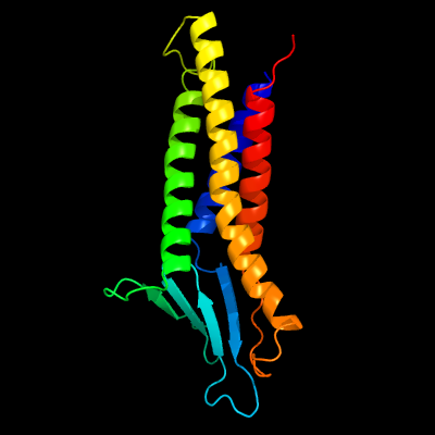
| c6ov2A_ | 3.20 | PDB header: cell adhesion | Chain: A: PDB Molecule: claudin-9; |
| Added to library: Sat Sep 7 08:24:12 2019 | |
| Links to external resources | |
|---|---|
| 1 | . | . | . | . | . | . | . | . | 10 | . | . | . | . | . | . | . | . | . | 20 | . | . | . | . | . | . | . | . | . | 30 | . | . | . | . | . | . | . | . | . | 40 | . | . | . | . | . | . | . | . | . | 50 | . | . | . | . | . | . | . | . | . | 60 | . | . | . | . | . | . | . | . | . | 70 | |
| Sequence | G | L | E | L | L | G | M | T | L | A | V | L | G | W | L | G | T | L | V | S | C | A | L | P | L | W | K | V | T | A | F | I | G | N | S | I | V | V | A | Q | V | V | W | E | G | L | W | M | S | C | V | V | Q | S | T | G | Q | M | Q | C | K | V | Y | D | S | L | L | A | L | P |
| Predicted secondary structure |  | | | | | | | | | | | | | | | | | | | |  | | | |  | | | | | | | | | | | | | | | | | | | | ||||||||||||||||||||||||||
| SS confidence | ||||||||||||||||||||||||||||||||||||||||||||||||||||||||||||||||||||||
| Known secondary structure (DSSP) | | | | | | | | | | | | | | | | | | | | | T | S | S | S | | | | | | S | T | T | S | S | | | | | | S | S | | | | | | S | S | S | S | | | | | S | S | S | . | . | . | . | . | . | . | . | . | 80 | . | . | . | . | . | . | . | . | . | 90 | . | . | . | . | . | . | . | . | . | 100 | . | . | . | . | . | . | . | . | . | 110 | . | . | . | . | . | . | . | . | . | 120 | . | . | . | . | . | . | . | . | . | 130 | . | . | . | . | . | . | . | . | . | 140 |
| Sequence | Q | D | L | Q | A | A | R | A | L | C | V | I | A | L | L | L | A | L | L | G | L | L | V | A | I | T | G | A | Q | C | T | T | C | V | E | D | E | G | A | K | A | R | I | V | L | T | A | G | V | I | L | L | L | A | G | I | L | V | L | I | P | V | C | W | T | A | H | A | I | I |
| Predicted secondary structure | | | | | | | | | | | | | | | | | | | | | | | | | | | | | | | | | | | | | | | | | | | | | | | | | | | | | | | | | | | | | | | | | | |||||
| SS confidence | ||||||||||||||||||||||||||||||||||||||||||||||||||||||||||||||||||||||
| Known secondary structure (DSSP) | | | | | | | | | | | | | | | | | | | | | | | | | T | T | S | S | T | T | S | S | | | | | | | | | | | | | | | | | | | | | | | | | | | | | | | | | | | . | . | . | . | . | . | . | . | . | 150 | . | . | . | . | . | . | . | . | . | 160 | . | . | . | . | . | . | . | . | . | 170 | . | . | . | . | . | . | . | . | . | 180 | . | . |
| Sequence | Q | D | F | Y | N | P | L | V | A | E | A | L | K | R | E | L | G | A | S | L | Y | L | G | W | A | A | A | A | L | L | M | L | G | G | G | L | L | C | C | T | C | P | ||||||||||||||||||||||||||||
| Predicted secondary structure | | | | | | | | | | | | | | | | | | | | | | | | | | | | | | | | |||||||||||||||||||||||||||||||||||||||
| SS confidence | ||||||||||||||||||||||||||||||||||||||||||||||||||||||||||||||||||||||
| Known secondary structure (DSSP) | G | G | G | G | T | T | S | S | S | B | B | | | | | | | | | | | | | | | | | | | | | |
| Download: | PDB structure | FASTA sequence |
Phyre is now FREE for commercial users! All images and data generated by Phyre2 are free to use in any publication with acknowledgement Accessibility Statement
| ||||||||||||||||||||||||