|
| ||||||
| Protein Homology/analogY Recognition Engine V 2.2 | |||||||
|
|
|
| ||||||
| Protein Homology/analogY Recognition Engine V 2.2 | |||||||
|
| Fold library id | PDB Header | Molecule | Title |
|---|---|---|---|
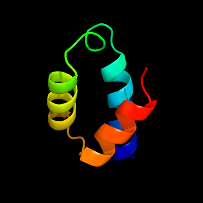
| c5aonB_ | PDB header: signaling protein | Chain: B: PDB Molecule: peroxin 14; | PDBTitle: crystal structure of the conserved n-terminal domain of2 pex14 from trypanosoma brucei |
| Added to library: Sat Dec 26 08:14:32 2015 | |
| Links to external resources | |
|---|---|
| 1 | . | . | . | . | . | . | . | . | 10 | . | . | . | . | . | . | . | . | . | 20 | . | . | . | . | . | . | . | . | . | 30 | . | . | . | . | . | . | . | . | . | 40 | . | . | . | . | . | . | . | . | . | |
| Sequence | S | E | R | E | K | R | V | S | N | A | V | E | F | L | L | D | S | R | V | R | R | T | P | T | S | S | K | V | H | F | L | K | S | K | G | L | S | A | E | E | I | C | E | A | F | T | K | V | G |
| Predicted secondary structure |  | | | | | | | | | | | | | | | | | | | | | | | | | | | | | | | | | | | | | ||||||||||||
| SS confidence | |||||||||||||||||||||||||||||||||||||||||||||||||
| Known secondary structure (DSSP) | | | | | | | | | | | | | | T | S | T | T | T | T | T | S | | | | | | | | | | | T | T | | | | | | | | | | T |
| Download: | PDB structure | FASTA sequence |
Phyre is now FREE for commercial users! All images and data generated by Phyre2 are free to use in any publication with acknowledgement Accessibility Statement
| ||||||||||||||||||||||||