|
| ||||||
| Protein Homology/analogY Recognition Engine V 2.2 | |||||||
|
|
|
| ||||||
| Protein Homology/analogY Recognition Engine V 2.2 | |||||||
|
| Fold library id | PDB Header | Molecule | Title |
|---|---|---|---|
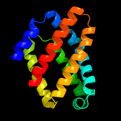
| c4zvbD_ | PDB header: signaling protein | Chain: D: PDB Molecule: diguanylate cyclase dosc; | PDBTitle: crystal structure of globin domain of the e. coli dosc - form ii2 (ferrous) |
| Added to library: Sat Nov 14 08:17:35 2015 | |
| Links to external resources | |
|---|---|
| 1 | . | . | . | . | . | . | . | . | 10 | . | . | . | . | . | . | . | . | . | 20 | . | . | . | . | . | . | . | . | . | 30 | . | . | . | . | . | . | . | . | . | 40 | . | . | . | . | . | . | . | . | . | 50 | . | . | . | . | . | . | . | . | . | 60 | . | . | . | . | . | . | . | . | . | 70 | |
| Sequence | K | R | M | K | D | E | W | T | G | L | V | E | Q | A | D | P | P | I | R | A | K | A | A | E | I | A | V | A | H | A | H | Y | L | S | I | E | F | Y | R | I | V | R | I | D | P | H | A | E | E | F | L | S | N | E | Q | V | E | R | Q | L | K | S | A | M | E | R | W | I | I | N |
| Predicted secondary structure |  | | | | | | | | | | | | | | | | | | | | | | | | | | | | | | | | | | | | | | | | | | | | | | | | | | | | | | | | | | | | | |||||||||
| SS confidence | ||||||||||||||||||||||||||||||||||||||||||||||||||||||||||||||||||||||
| Known secondary structure (DSSP) | | | | | | | | | | | | S | S | | | | | | | | | | | | | | | | | | | | | | | | | | | T | T | S | | | | | T | T | S | S | | | | | | | | | | | | | | | | | | . | . | . | . | . | . | . | . | . | 80 | . | . | . | . | . | . | . | . | . | 90 | . | . | . | . | . | . | . | . | . | 100 | . | . | . | . | . | . | . | . | . | 110 | . | . | . | . | . | . | . | . | . | 120 | . | . | . | . | . | . | . | . | . | 130 | . | . | . | . | . | . | . | . | . | 140 |
| Sequence | V | L | S | A | Q | V | D | D | V | E | R | L | I | Q | I | Q | H | T | V | A | E | V | H | A | R | I | G | I | P | V | E | I | V | E | M | G | F | R | V | L | K | K | I | L | Y | P | V | I | F | S | S | D | Y | S | A | A | E | K | L | Q | V | Y | H | F | S | I | N | S | I | D |
| Predicted secondary structure | | | | | | | | | | | | | | | | | | | | | | | | | | | | | | | | | | | | | | | | | | | | | | | | | | | | | | | | | ||||||||||||||
| SS confidence | ||||||||||||||||||||||||||||||||||||||||||||||||||||||||||||||||||||||
| Known secondary structure (DSSP) | | | T | G | G | G | | | | | | | | | | | | | | | | | | | T | | | | | | | | | | | | | | | | | | | | | T | S | S | S | | | | | | | | | | | | | | | | | . | . | . | . | . | . | . | . | . |
| Sequence | I | A | M | E | V | M | T | R | A | |||||||||||||||||||||||||||||||||||||||||||||||||||||||||||||
| Predicted secondary structure | | | | | | | | |||||||||||||||||||||||||||||||||||||||||||||||||||||||||||||||
| SS confidence | ||||||||||||||||||||||||||||||||||||||||||||||||||||||||||||||||||||||
| Known secondary structure (DSSP) | | | | | | | | T |
| Download: | PDB structure | FASTA sequence |
Phyre is now FREE for commercial users! All images and data generated by Phyre2 are free to use in any publication with acknowledgement Accessibility Statement
| ||||||||||||||||||||||||