|
| ||||||
| Protein Homology/analogY Recognition Engine V 2.2 | |||||||
|
|
|
| ||||||
| Protein Homology/analogY Recognition Engine V 2.2 | |||||||
|
| Fold library id | PDB Header | Molecule | Title |
|---|---|---|---|
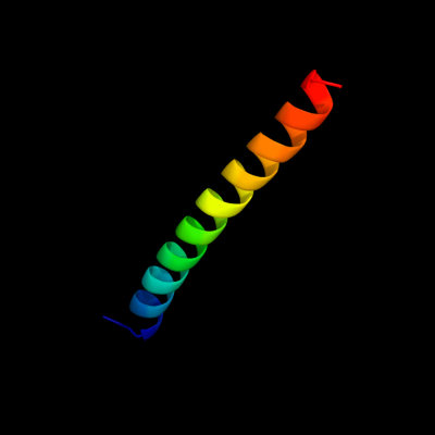
| c4ds7G_ | PDB header: protein binding | Chain: G: PDB Molecule: spindle pole body component 110; | PDBTitle: crystal structure of yeast calmodulin bound to the c-terminal fragment2 of spindle pole body protein spc110 |
| Added to library: Thu Jan 10 04:24:47 2013 | |
| Links to external resources | |
|---|---|
| 1 | . | . | . | . | . | . | . | . | 10 | . | . | . | . | . | . | . | . | . | 20 | . | . | . | . | . | . | . | . | . | 30 | . | . | . | . | . | . | |
| Sequence | L | S | F | K | T | V | A | L | L | V | L | A | C | V | R | M | K | R | I | A | F | Y | R | R | S | D | D | N | R | L | R | I | L | R | D | R |
| Predicted secondary structure |  | | | | | | | | | | | | | | | | | |  | | |  | | | | | | | ||||||||
| SS confidence | ||||||||||||||||||||||||||||||||||||
| Known secondary structure (DSSP) | | | | | | | | | | | | | | | | | | | | | | | | | | | | | | | | | |
| Download: | PDB structure | FASTA sequence |
Phyre is now FREE for commercial users! All images and data generated by Phyre2 are free to use in any publication with acknowledgement Accessibility Statement
| ||||||||||||||||||||||||