|
| ||||||
| Protein Homology/analogY Recognition Engine V 2.2 | |||||||
|
|
|
| ||||||
| Protein Homology/analogY Recognition Engine V 2.2 | |||||||
|
| Fold library id | PDB Header | Molecule | Title |
|---|---|---|---|
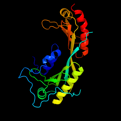
| c4b0aA_ | PDB header: transcription | Chain: A: PDB Molecule: transcription initiation factor tfiid subunit 1, linker, | PDBTitle: the high-resolution structure of ytbp-ytaf1 identifies2 conserved and competing interaction surfaces in3 transcriptional activation |
| Added to library: Sat Jul 6 08:26:47 2013 | |
| Links to external resources | |
|---|---|
| 1 | . | . | . | . | . | . | . | . | 10 | . | . | . | . | . | . | . | . | . | 20 | . | . | . | . | . | . | . | . | . | 30 | . | . | . | . | . | . | . | . | . | 40 | . | . | . | . | . | . | . | . | . | 50 | . | . | . | . | . | . | . | . | . | 60 | . | . | . | . | . | . | . | . | . | 70 | |
| Sequence | K | T | N | L | A | N | E | D | E | A | Y | E | A | I | F | G | G | E | F | G | S | L | E | I | G | S | Y | I | G | G | D | E | A | R | N | S | K | D | Y | T | E | H | L | P | D | A | V | D | F | E | D | E | D | E | L | A | D | D | G | I | V | P | T | L | Q | N | I | V | A | T |
| Predicted secondary structure |  | | | | | | | |  | |  | | | | | | | | | | | | | | | |||||||||||||||||||||||||||||||||||||||||||||
| SS confidence | ||||||||||||||||||||||||||||||||||||||||||||||||||||||||||||||||||||||
| Known secondary structure (DSSP) | T | T | S | | | | | | | | | | S | S | S | G | G | G | G | G | G | G | G | S | T | T | S | S | T | T | B | T | T | | | | | | | | | . | . | . | . | . | . | . | . | . | 80 | . | . | . | . | . | . | . | . | . | 90 | . | . | . | . | . | . | . | . | . | 100 | . | . | . | . | . | . | . | . | . | 110 | . | . | . | . | . | . | . | . | . | 120 | . | . | . | . | . | . | . | . | . | 130 | . | . | . | . | . | . | . | . | . | 140 |
| Sequence | V | T | L | G | C | R | L | D | L | K | T | V | A | L | H | A | R | N | A | E | Y | N | P | K | R | F | A | A | V | I | M | R | I | R | E | P | K | T | T | A | L | I | F | A | S | G | K | M | V | V | T | G | A | K | S | E | D | D | S | K | L | A | S | R | K | Y | A | R | I | I |
| Predicted secondary structure | | | | | | | | | | | | | | | | | | | | | | | | | | | | | | | | | | | | | | | | | | | | | | | | |||||||||||||||||||||||
| SS confidence | ||||||||||||||||||||||||||||||||||||||||||||||||||||||||||||||||||||||
| Known secondary structure (DSSP) | | | S | S | | | | | | | | B | T | T | | | | T | T | T | S | S | | | | | | T | T | T | T | | | | | | T | T | S | | | | | | | | S | S | | | | | | | | | | | | | | | | . | . | . | . | . | . | . | . | . | 150 | . | . | . | . | . | . | . | . | . | 160 | . | . | . | . | . | . | . | . | . | 170 | . | . | . | . | . | . | . | . | . | 180 | . | . | . | . | . | . | . | . | . | 190 | . | . | . | . | . | . | . | . | . | 200 | . | . | . | . | . | . | . | . | . | 210 |
| Sequence | Q | K | I | G | F | A | A | K | F | T | D | F | K | I | Q | N | I | V | G | S | C | D | V | K | F | P | I | R | L | E | G | L | A | F | S | H | G | T | F | S | S | Y | E | P | E | L | F | P | G | L | I | Y | R | M | V | K | P | K | I | V | L | L | I | F | V | S | G | K | I | V |
| Predicted secondary structure | | | | | | | | | | | | | | | | | | | | | | | | | | | | | | | | | | | | |||||||||||||||||||||||||||||||||||
| SS confidence | ||||||||||||||||||||||||||||||||||||||||||||||||||||||||||||||||||||||
| Known secondary structure (DSSP) | | | | T | | | | | | | | | | | | | | S | S | B | | | | | | | | T | T | T | T | | | T | T | T | S | S | | | | | S | S | S | | | | | T | T | S | | | | . | . | . | . | . | . | . | . | . | 220 | . | . | . | . | . | . | . | . | . | 230 | . | . | . | . | . | . | . |
| Sequence | L | T | G | A | K | Q | R | E | E | I | Y | Q | A | F | E | A | I | Y | P | V | L | S | E | F | R | K | M | |||||||||||||||||||||||||||||||||||||||||||
| Predicted secondary structure | | | | | | | | | | | | | | | | | | | | | | | ||||||||||||||||||||||||||||||||||||||||||||||||
| SS confidence | ||||||||||||||||||||||||||||||||||||||||||||||||||||||||||||||||||||||
| Known secondary structure (DSSP) | | | | | S | S | | | | | | | | | | | | | | | | | | T | B |
| Download: | PDB structure | FASTA sequence |
Phyre is now FREE for commercial users! All images and data generated by Phyre2 are free to use in any publication with acknowledgement Accessibility Statement
| ||||||||||||||||||||||||