|
| ||||||
| Protein Homology/analogY Recognition Engine V 2.2 | |||||||
|
|
|
| ||||||
| Protein Homology/analogY Recognition Engine V 2.2 | |||||||
|
| Fold library id | PDB Header | Molecule | Title |
|---|---|---|---|
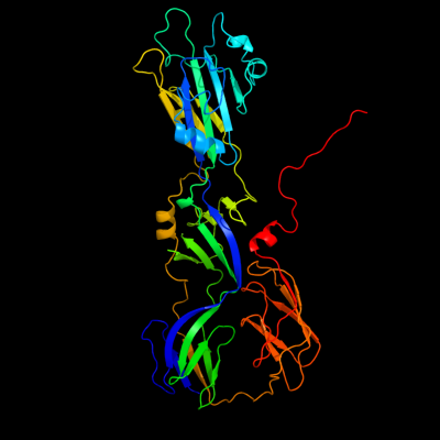
| c4adjB_ | PDB header: viral protein | Chain: B: PDB Molecule: e1 envelope glycoprotein; | PDBTitle: crystal structure of the rubella virus glycoprotein e1 in its2 post-fusion form crystallized in presence of 1mm of calcium acetate |
| Added to library: Mon Jan 14 11:28:53 2013 | |
| Links to external resources | |
|---|---|
| 1 | . | . | . | . | . | . | . | . | 10 | . | . | . | . | . | . | . | . | . | 20 | . | . | . | . | . | . | . | . | . | 30 | . | . | . | . | . | . | . | . | . | 40 | . | . | . | . | . | . | . | . | . | 50 | . | . | . | . | . | . | . | . | . | 60 | . | . | . | . | . | . | . | . | . | 70 | |
| Sequence | E | A | F | T | Y | L | C | T | A | P | G | C | A | T | Q | T | P | V | P | V | R | L | A | G | V | R | F | E | S | K | I | V | D | G | G | C | F | A | P | W | D | L | E | A | T | G | A | C | I | C | E | I | P | T | D | V | S | C | E | G | L | G | A | W | V | P | T | A | P | C |
| Predicted secondary structure |  | | | |  | | | | | | | | | | | | | | | | | |  | |||||||||||||||||||||||||||||||||||||||||||||||
| SS confidence | ||||||||||||||||||||||||||||||||||||||||||||||||||||||||||||||||||||||
| Known secondary structure (DSSP) | | | | | | S | T | T | | | S | S | S | | | | | | | | | | | | | | | | | | | | | | | | | | | | S | S | S | S | T | T | T | T | T | T | T | S | | | | | . | . | . | . | . | . | . | . | . | 80 | . | . | . | . | . | . | . | . | . | 90 | . | . | . | . | . | . | . | . | . | 100 | . | . | . | . | . | . | . | . | . | 110 | . | . | . | . | . | . | . | . | . | 120 | . | . | . | . | . | . | . | . | . | 130 | . | . | . | . | . | . | . | . | . | 140 |
| Sequence | A | R | I | W | N | G | T | Q | R | A | C | T | F | W | A | V | N | A | Y | S | S | G | G | Y | A | Q | L | A | S | Y | F | N | P | G | G | S | Y | Y | K | Q | Y | H | P | T | A | C | E | V | E | P | A | F | G | H | S | D | A | A | C | W | G | F | P | T | D | T | V | M | S | V |
| Predicted secondary structure | | | | | | | | | | | | | | | | | | | | | | | | | | | | | | | | | | | | | | |||||||||||||||||||||||||||||||||
| SS confidence | ||||||||||||||||||||||||||||||||||||||||||||||||||||||||||||||||||||||
| Known secondary structure (DSSP) | | | | | T | S | | | | | | | | | | | | B | S | S | S | S | S | B | G | G | G | T | T | | | | | | | S | S | S | | | | | T | T | S | T | T | S | S | S | | | | . | . | . | . | . | . | . | . | . | 150 | . | . | . | . | . | . | . | . | . | 160 | . | . | . | . | . | . | . | . | . | 170 | . | . | . | . | . | . | . | . | . | 180 | . | . | . | . | . | . | . | . | . | 190 | . | . | . | . | . | . | . | . | . | 200 | . | . | . | . | . | . | . | . | . | 210 |
| Sequence | F | A | L | A | S | Y | V | Q | H | P | H | K | T | V | R | V | K | F | H | T | E | T | R | T | V | W | Q | L | S | V | A | G | V | S | C | N | V | T | T | E | H | P | F | C | N | T | P | H | G | Q | L | E | V | Q | V | P | P | D | P | G | D | L | V | E | Y | I | M | N | N | Q |
| Predicted secondary structure | | | | | | | | | | | | | | | | | | | | | | | | | | | | | | | | | | | | | | | | | | | | | | | ||||||||||||||||||||||||
| SS confidence | ||||||||||||||||||||||||||||||||||||||||||||||||||||||||||||||||||||||
| Known secondary structure (DSSP) | | | | | | | G | G | G | | | | | | | | | | | | | | | | | | T | T | | | | | | S | S | | | | S | S | S | | | | | | | | | | | | . | . | . | . | . | . | . | . | . | 220 | . | . | . | . | . | . | . | . | . | 230 | . | . | . | . | . | . | . | . | . | 240 | . | . | . | . | . | . | . | . | . | 250 | . | . | . | . | . | . | . | . | . | 260 | . | . | . | . | . | . | . | . | . | 270 | . | . | . | . | . | . | . | . | . | 280 |
| Sequence | Q | S | R | W | G | L | G | S | P | N | C | H | G | P | D | W | A | S | P | V | C | Q | R | H | S | P | D | C | S | R | L | V | G | A | T | P | E | R | P | R | L | R | L | V | D | A | D | D | P | L | L | R | T | A | P | G | P | G | E | V | W | V | T | P | V | I | G | S | Q | A |
| Predicted secondary structure | | | | | | | | | | | | | | | | | | | | | | | | | | | | |||||||||||||||||||||||||||||||||||||||||||
| SS confidence | ||||||||||||||||||||||||||||||||||||||||||||||||||||||||||||||||||||||
| Known secondary structure (DSSP) | | | | | | | S | S | T | T | S | T | T | T | T | G | G | G | S | | | | T | T | S | S | S | B | S | | | | | T | T | G | G | G | G | S | T | T | | | | | | | | | S | S | T | . | . | . | . | . | . | . | . | . | 290 | . | . | . | . | . | . | . | . | . | 300 | . | . | . | . | . | . | . | . | . | 310 | . | . | . | . | . | . | . | . | . | 320 | . | . | . | . | . | . | . | . | . | 330 | . | . | . | . | . | . | . | . | . | 340 | . | . | . | . | . | . | . | . | . | 350 |
| Sequence | R | K | C | G | L | H | I | R | A | G | P | Y | G | H | A | T | V | E | M | P | E | W | I | H | A | H | T | T | S | D | P | W | H | P | P | G | P | L | G | L | K | F | K | T | V | R | P | V | A | L | P | R | A | L | A | P | P | R | N | V | R | V | T | G | C | Y | Q | C | G | T |
| Predicted secondary structure | | | | | | | | | | | | | | | | | | | | | | | | | | | | | ||||||||||||||||||||||||||||||||||||||||||
| SS confidence | ||||||||||||||||||||||||||||||||||||||||||||||||||||||||||||||||||||||
| Known secondary structure (DSSP) | T | | | | | | | | G | G | G | S | | | | | | | | | B | S | S | | | | | | | B | S | | | | | | | | | S | S | S | S | S | . | . | . | . | . | . | . | . | . | 360 | . | . | . | . | . | . | . | . | . | 370 | . | . | . | . | . | . | . | . | . | 380 | . | . | . | . | . | . | . | . | . | 390 | . | . | . | . | . | . | . | . | . | 400 | . | . | . | . | . | . | . | . | . | 410 | . | . | . | . | . | . | . | . | . | 420 |
| Sequence | P | A | L | V | E | G | L | A | P | G | G | G | N | C | H | L | T | V | N | G | E | D | V | G | A | F | P | P | G | K | F | V | T | A | A | L | L | N | T | P | P | P | Y | Q | V | S | C | G | G | E | S | D | R | A | S | A | R | V | I | D | P | A | A | Q | S | F | T | G | V | V |
| Predicted secondary structure | | | | | | | | | | | | | | | | | | | | | | | | | | | | | | | | | | | ||||||||||||||||||||||||||||||||||||
| SS confidence | ||||||||||||||||||||||||||||||||||||||||||||||||||||||||||||||||||||||
| Known secondary structure (DSSP) | | | | | | | | S | S | S | B | | | | | | T | T | | | | | | | S | | | | | | | | S | S | S | | | | S | S | S | S | | | | | | | | | | | | | | T | T | T | . | . | . | . | . | . | . | . | . | 430 | . | |
| Sequence | Y | G | T | H | T | T | A | V | S | E | T | |||||||||||||||||||||||||||||||||||||||||||||||||||||||||||
| Predicted secondary structure | | | | | | | | | ||||||||||||||||||||||||||||||||||||||||||||||||||||||||||||||
| SS confidence | ||||||||||||||||||||||||||||||||||||||||||||||||||||||||||||||||||||||
| Known secondary structure (DSSP) | S |
| Download: | PDB structure | FASTA sequence |
Phyre is now FREE for commercial users! All images and data generated by Phyre2 are free to use in any publication with acknowledgement Accessibility Statement
| ||||||||||||||||||||||||