|
| ||||||
| Protein Homology/analogY Recognition Engine V 2.2 | |||||||
|
|
|
| ||||||
| Protein Homology/analogY Recognition Engine V 2.2 | |||||||
|
| Fold library id | PDB Header | Molecule | Title |
|---|---|---|---|
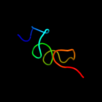
| c3v28U_ | PDB header: ribosome | Chain: U: PDB Molecule: 30s ribosomal protein thx; | PDBTitle: crystal structure of hpf bound to the 70s ribosome. this pdb entry2 contains coordinates for the 30s subunit with bound hpf of the 2nd3 ribosome in the asu |
| Added to library: Wed Jan 9 19:59:44 2013 | |
| Links to external resources | |
|---|---|
| 1 | . | . | . | . | . | . | . | . | 10 | . | . | . | . | . | . | . | . | . | 20 | . | . | . | . | . | . | |
| Sequence | G | K | G | D | R | R | T | R | R | G | K | I | W | R | G | T | Y | G | K | Y | R | P | R | X | X | X |
| Predicted secondary structure |  | |  | |||||||||||||||||||||||
| SS confidence | ||||||||||||||||||||||||||
| Known secondary structure (DSSP) | S | S | S | G | G | G | T | T | T | T | T | S | S | S | S | - | - | - |
| Download: | PDB structure | FASTA sequence |
Phyre is now FREE for commercial users! All images and data generated by Phyre2 are free to use in any publication with acknowledgement Accessibility Statement
| ||||||||||||||||||||||||