|
| ||||||
| Protein Homology/analogY Recognition Engine V 2.2 | |||||||
|
|
|
| ||||||
| Protein Homology/analogY Recognition Engine V 2.2 | |||||||
|
| Fold library id | PDB Header | Molecule | Title |
|---|---|---|---|
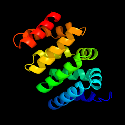
| c3sl9G_ | PDB header: signaling protein, protein binding | Chain: G: PDB Molecule: catenin beta-1; | PDBTitle: x-ray structure of beta catenin in complex with bcl9 |
| Added to library: Sat Mar 3 11:45:41 2012 | |
| Links to external resources | |
|---|---|
| 1 | . | . | . | . | . | . | . | . | 10 | . | . | . | . | . | . | . | . | . | 20 | . | . | . | . | . | . | . | . | . | 30 | . | . | . | . | . | . | . | . | . | 40 | . | . | . | . | . | . | . | . | . | 50 | . | . | . | . | . | . | . | . | . | 60 | . | . | . | . | . | . | . | . | . | 70 | |
| Sequence | L | A | T | R | A | I | P | E | L | T | K | L | L | N | D | E | D | Q | V | V | V | N | K | A | A | V | M | V | H | Q | L | S | K | K | E | A | S | R | H | A | I | M | R | S | P | Q | M | V | S | A | I | V | R | T | M | Q | N | T | N | D | V | E | T | A | R | C | T | A | G | T |
| Predicted secondary structure |  | | | | | | | | | | | | | | | | | | | | | | | | | | | | | | | | | | | | | | | | | | | | | | | | | | ||||||||||||||||||||
| SS confidence | ||||||||||||||||||||||||||||||||||||||||||||||||||||||||||||||||||||||
| Known secondary structure (DSSP) | | | | | | | | T | S | | | | | | | | | | | | | | | T | T | S | | | | | | | | | T | | | | | | | | | | | | | | | | | | | | | | | | . | . | . | . | . | . | . | . | . | 80 | . | . | . | . | . | . | . | . | . | 90 | . | . | . | . | . | . | . | . | . | 100 | . | . | . | . | . | . | . | . | . | 110 | . | . | . | . | . | . | . | . | . | 120 | . | . | . | . | . | . | . | . | . | 130 | . | . | . | . | . | . | . | . | . | 140 |
| Sequence | L | H | N | L | S | H | H | R | E | G | L | L | A | I | F | K | S | G | G | I | P | A | L | V | K | M | L | G | S | P | V | D | S | V | L | F | Y | A | I | T | T | L | H | N | L | L | L | H | Q | E | G | A | K | M | A | V | R | L | A | G | G | L | Q | K | M | V | A | L | L | N |
| Predicted secondary structure | | | | | | | | | | | | | | | | | | | | | | | | | | | | | | | | | | | | | | | | | | | | | | | | | ||||||||||||||||||||||
| SS confidence | ||||||||||||||||||||||||||||||||||||||||||||||||||||||||||||||||||||||
| Known secondary structure (DSSP) | | | | | | T | S | | | | | | | | | | T | T | | | | | | | | | T | T | S | | | | | | | | | | | | | | | | | | T | T | | | | | | | | T | T | | | | | | | | | T | T | . | . | . | . | . | . | . | . | . | 150 | . | . | . | . | . | . | . | . |
| Sequence | K | T | N | V | K | F | L | A | I | T | T | D | C | L | Q | I | L | A | ||||||||||||||||||||||||||||||||||||||||||||||||||||
| Predicted secondary structure | | | | | | | | | | | | | | | ||||||||||||||||||||||||||||||||||||||||||||||||||||||||
| SS confidence | ||||||||||||||||||||||||||||||||||||||||||||||||||||||||||||||||||||||
| Known secondary structure (DSSP) | S | S | | | | | | | | | | | | | | |
| Download: | PDB structure | FASTA sequence |
Phyre is now FREE for commercial users! All images and data generated by Phyre2 are free to use in any publication with acknowledgement Accessibility Statement
| ||||||||||||||||||||||||