|
| ||||||
| Protein Homology/analogY Recognition Engine V 2.2 | |||||||
|
|
|
| ||||||
| Protein Homology/analogY Recognition Engine V 2.2 | |||||||
|
| Fold library id | PDB Header | Molecule | Title |
|---|---|---|---|
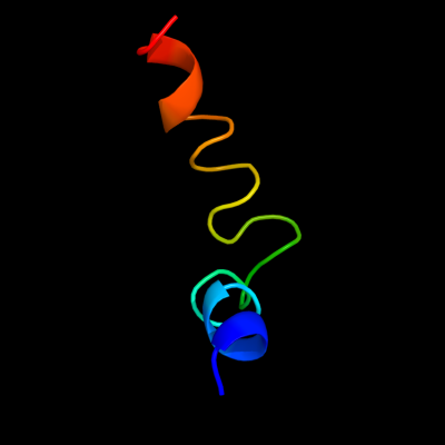
| c3i8iJ_ | PDB header: ribosome | Chain: J: PDB Molecule: 50s ribosomal protein l7/l12; | PDBTitle: elongation complex of the 70s ribosome with three trnas and mrna. this2 entry 3i8i contains 50s ribosomal subnit. the 30s ribosomal subunit3 can be found in pdb entry 3i8h. molecule a in the same asymmetric4 unit is deposited as 3i8f (50s) and 3i8g (30s). |
| Added to library: Thu Jan 10 01:08:57 2013 | |
| Links to external resources | |
|---|---|
| 1 | . | . | . | . | . | . | . | . | 10 | . | . | . | . | . | . | . | . | . | 20 | . | . | . | . | . | . | . | . | . | 30 | |
| Sequence | M | A | L | D | I | E | R | I | K | E | E | L | S | Q | A | T | V | L | E | L | K | Q | L | I | D | A | L | K | E | A |
| Predicted secondary structure |  | | | | | | | | | | | | | | | | | | | | | | | | ||||||
| SS confidence | ||||||||||||||||||||||||||||||
| Known secondary structure (DSSP) | | | | | | T | T | T | S | S | S | S | S | S | S | S | T | T | S | T | T | T | G | G | G | T |
| Download: | PDB structure | FASTA sequence |
Phyre is now FREE for commercial users! All images and data generated by Phyre2 are free to use in any publication with acknowledgement Accessibility Statement
| ||||||||||||||||||||||||