|
| ||||||
| Protein Homology/analogY Recognition Engine V 2.2 | |||||||
|
|
|
| ||||||
| Protein Homology/analogY Recognition Engine V 2.2 | |||||||
|
| Fold library id | PDB Header | Molecule | Title |
|---|---|---|---|
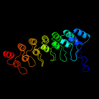
| c3d9hA_ | PDB header: structural protein, protein binding | Chain: A: PDB Molecule: cdna flj77766, highly similar to homo sapiens | PDBTitle: crystal structure of the splice variant of human asb92 (hasb9-2), an ankyrin repeat protein |
| Added to library: Sun Aug 29 01:02:00 2010 | |
| Links to external resources | |
|---|---|
| 1 | . | . | . | . | . | . | . | . | 10 | . | . | . | . | . | . | . | . | . | 20 | . | . | . | . | . | . | . | . | . | 30 | . | . | . | . | . | . | . | . | . | 40 | . | . | . | . | . | . | . | . | . | 50 | . | . | . | . | . | . | . | . | . | 60 | . | . | . | . | . | . | . | . | . | 70 | |
| Sequence | F | P | G | I | R | L | L | S | N | P | L | M | G | D | A | V | S | D | W | S | P | M | H | E | A | A | I | H | G | H | Q | L | S | L | R | N | L | I | S | Q | G | W | A | V | N | I | I | T | A | D | H | V | S | P | L | H | E | A | C | L | G | G | H | L | S | C | V | K | I | L |
| Predicted secondary structure |  | | | | | | | | | | | | | | | | ||||||||||||||||||||||||||||||||||||||||||||||||||||||
| SS confidence | ||||||||||||||||||||||||||||||||||||||||||||||||||||||||||||||||||||||
| Known secondary structure (DSSP) | S |  |  | T | T | S | S | S | S | | | | | | | | T | T | | | | | | | | | | T | T | S | | | T | T | | | | | | | | T | T | | | | | | | | . | . | . | . | . | . | . | . | . | 80 | . | . | . | . | . | . | . | . | . | 90 | . | . | . | . | . | . | . | . | . | 100 | . | . | . | . | . | . | . | . | . | 110 | . | . | . | . | . | . | . | . | . | 120 | . | . | . | . | . | . | . | . | . | 130 | . | . | . | . | . | . | . | . | . | 140 |
| Sequence | L | K | H | G | A | Q | V | N | G | V | T | A | D | W | H | T | P | L | F | N | A | C | V | S | G | S | W | D | C | V | N | L | L | L | Q | H | G | A | S | V | Q | P | E | S | D | L | A | S | P | I | H | E | A | A | R | R | G | H | V | E | C | V | N | S | L | I | A | Y | G | G |
| Predicted secondary structure | ||||||||||||||||||||||||||||||||||||||||||||||||||||||||||||||||||||||
| SS confidence | ||||||||||||||||||||||||||||||||||||||||||||||||||||||||||||||||||||||
| Known secondary structure (DSSP) | | | T | T | S | S | T | T | | | | | | | | | T | | | | | | | | | | T | T | S | S | S | T | T | S | | | | | | | | | T | | | | | | | | | | T | T | . | . | . | . | . | . | . | . | . | 150 | . | . | . | . | . | . | . | . | . | 160 | . | . | . | . | . | . | . | . | . | 170 | . | . | . | . | . | . | . | . | . | 180 | . | . | . | . | . | . | . | . | . | 190 | . | . | . | . | . | . | . | . | . | 200 | . | . | . | . | . | . | . | . | . | 210 |
| Sequence | N | I | D | H | K | I | S | H | L | G | T | P | L | Y | L | A | C | E | N | Q | Q | R | A | C | V | K | K | L | L | E | S | G | A | D | V | N | Q | G | K | G | Q | D | S | P | L | H | A | V | V | R | T | A | S | E | E | L | A | C | L | L | M | D | F | G | A | D | T | Q | A | K |
| Predicted secondary structure | | | | | | | | | | | | | | | ||||||||||||||||||||||||||||||||||||||||||||||||||||||||
| SS confidence | ||||||||||||||||||||||||||||||||||||||||||||||||||||||||||||||||||||||
| Known secondary structure (DSSP) | T | T | B | T | T | T | B | | | | | | | | T | T | | | | | | | | | | T | T | T | T | B | T | T | B | | | | | | | | T | T | | | | | | | | | | T | T | T | T | . | . | . | . | . | . | . | . | . | 220 | . | . | . | . | . | . | . | . | . | 230 | . | . | . | . | . |
| Sequence | N | A | E | G | K | R | P | V | E | L | V | P | P | E | S | P | L | A | Q | L | F | L | E | R | E | |||||||||||||||||||||||||||||||||||||||||||||
| Predicted secondary structure | | | | | | | | | | | | | | |||||||||||||||||||||||||||||||||||||||||||||||||||||||||
| SS confidence | ||||||||||||||||||||||||||||||||||||||||||||||||||||||||||||||||||||||
| Known secondary structure (DSSP) | T | T | S | G | G | G | G | S | T | T | S | | | | | | | | | |
| Download: | PDB structure | FASTA sequence |
Phyre is now FREE for commercial users! All images and data generated by Phyre2 are free to use in any publication with acknowledgement Accessibility Statement
| ||||||||||||||||||||||||