|
| ||||||
| Protein Homology/analogY Recognition Engine V 2.2 | |||||||
|
|
|
| ||||||
| Protein Homology/analogY Recognition Engine V 2.2 | |||||||
|
| Fold library id | PDB Header | Molecule | Title |
|---|---|---|---|
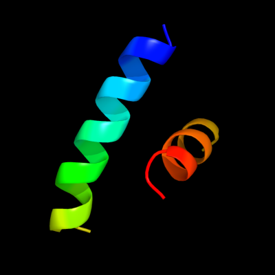
| c3d5dJ_ | PDB header: ribosome | Chain: J: PDB Molecule: 50s ribosomal protein l10; | PDBTitle: structural basis for translation termination on the 70s ribosome. this2 file contains the 50s subunit of the second 70s ribosome. the entire3 crystal structure contains two 70s ribosomes as described in remark4 400. |
| Added to library: Wed Jan 9 20:46:56 2013 | |
| Links to external resources | |
|---|---|
| 1 | . | . | . | . | . | . | . | . | 10 | . | . | . | . | . | . | . | . | . | 20 | . | . | . | . | . | . | . | . | . | 30 | . | . | |
| Sequence | N | K | R | N | V | E | L | L | A | T | L | K | E | N | L | E | R | A | Q | G | N | T | L | I | R | L | A | L | K | E | L | G |
| Predicted secondary structure |  | | | | | | | | | | | | | | | | | | | | | | | | | | ||||||
| SS confidence | ||||||||||||||||||||||||||||||||
| Known secondary structure (DSSP) | | | | | T | T | T | T | T | | | | | | | T | T | | | | | | | | | T | T |
| Download: | PDB structure | FASTA sequence |
Phyre is now FREE for commercial users! All images and data generated by Phyre2 are free to use in any publication with acknowledgement Accessibility Statement
| ||||||||||||||||||||||||