|
| ||||||
| Protein Homology/analogY Recognition Engine V 2.2 | |||||||
|
|
|
| ||||||
| Protein Homology/analogY Recognition Engine V 2.2 | |||||||
|
| Fold library id | PDB Header | Molecule | Title |
|---|---|---|---|
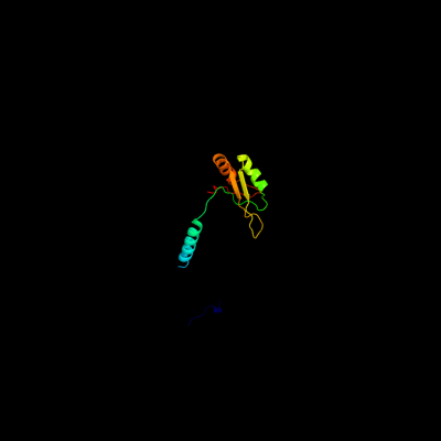
| c3cw18_ | PDB header: splicing | Chain: 8: PDB Molecule: u1 small nuclear ribonucleoprotein 70 kda; | PDB Fragment:nucleotides 57-82 absent, replaced with kissing |
| Added to library: Sat Aug 28 17:49:44 2010 | |
| Links to external resources | |
|---|---|
| 1 | . | . | . | . | . | . | . | . | 10 | . | . | . | . | . | . | . | . | . | 20 | . | . | . | . | . | . | . | . | . | 30 | . | . | . | . | . | . | . | . | . | 40 | . | . | . | . | . | . | . | . | . | 50 | . | . | . | . | . | . | . | . | . | 60 | . | . | . | . | . | . | . | . | . | 70 | |
| Sequence | L | L | A | L | F | A | P | R | D | P | I | P | Y | L | P | R | E | E | R | M | E | R | K | R | R | E | K | I | E | R | R | Q | Q | E | V | E | T | E | L | K | M | W | K | T | L | F | V | A | R | V | N | Y | D | T | T | E | S | K | L | R | R | E | F | E | V | Y | G | P | I | K |
| Predicted secondary structure |  | | | | | | | | | | |  | | |  | | | | | | | | | | | |||||||||||||||||||||||||||||||||||||||||||||
| SS confidence | ||||||||||||||||||||||||||||||||||||||||||||||||||||||||||||||||||||||
| Known secondary structure (DSSP) | T | T | T | S | S | | | | | | | | | | | | | | G | G | G | B | T | T | | | | | | | | | | T | T | S | | | . | . | . | . | . | . | . | . | . | 80 | . | . | . | . | . | . | . | . | . | 90 | . | . | . | . | . | . | . | . | . | 100 | . | . | . | . | . | . | . | . | . | 110 | . | . | . | . | . | . | . | . | . | 120 |
| Sequence | R | I | H | M | V | Y | S | K | R | S | G | K | P | R | G | Y | A | F | I | E | Y | E | H | E | R | D | M | H | S | A | Y | K | H | A | D | G | K | K | I | D | G | R | R | V | L | V | D | V | E | R | ||||||||||||||||||||
| Predicted secondary structure | | | | | | | | | | | | | | | | | | | | | | | | | | | | | | | | |||||||||||||||||||||||||||||||||||||||
| SS confidence | ||||||||||||||||||||||||||||||||||||||||||||||||||||||||||||||||||||||
| Known secondary structure (DSSP) | | | | | | | S | S | S | S | | | | | | | | | | S | S | | | | | | | | | | | | T | T | | | S | S | S | | | B |
| Download: | PDB structure | FASTA sequence |
Phyre is now FREE for commercial users! All images and data generated by Phyre2 are free to use in any publication with acknowledgement Accessibility Statement
| ||||||||||||||||||||||||